42 amoeba diagram labeled
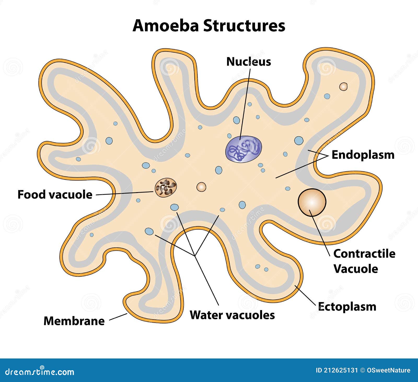
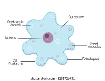
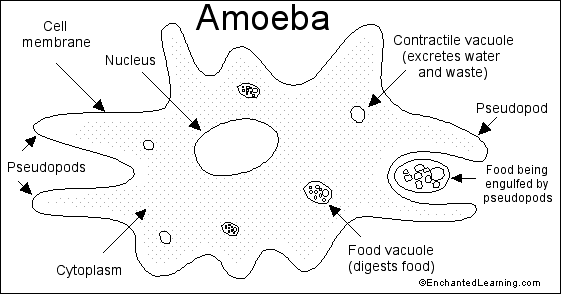
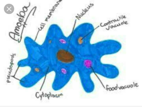
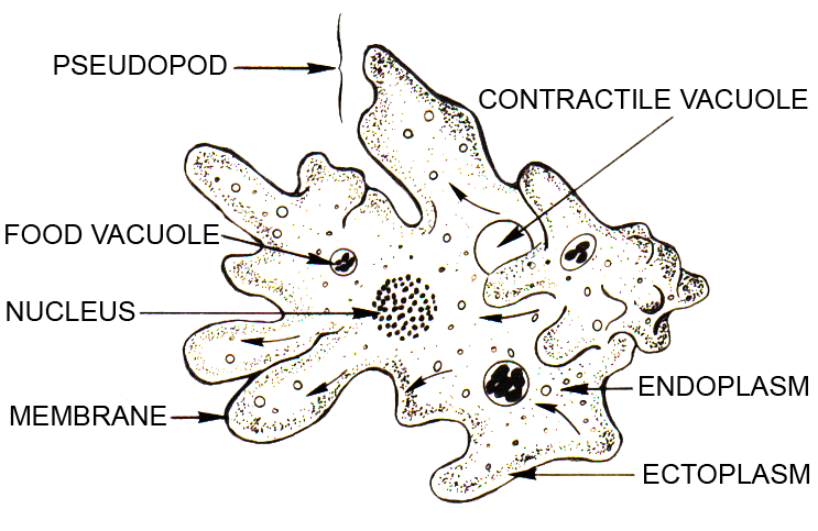
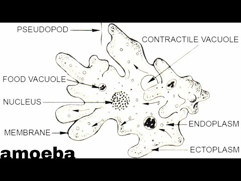
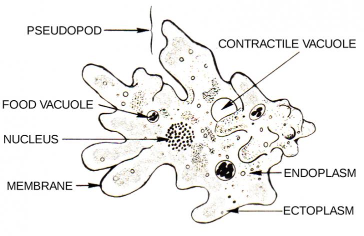
emcee101 Label Amoeba Diagram STUDY PLAY Cell Membrane The thin layer of protein and fat that surrounds the ameoba; it allows some substances to pass into the cell, and block other substances contractile vacoule Amoeba exhibits movement by the pseudopodia. It also helps in food capture. Like an ordinary cell the body of amoeba has 3 main parts: Plasma lemma or plasma membrane, Cytoplasm and nucleus. Plasma lemma is a very thin, delicate and elastic cell membrane of amoeba. It is composed of a double layer of lipid and protein molecules.
Download this Amoeba Labeled Vector Illustration Single Cell Animal Structure Scheme vector illustration now. And search more of iStock's library of royalty-free vector art that features Amoeba graphics available for quick and easy download.
Amoeba diagram labeled
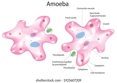
Amoeba labeled vector illustration. Educational single cell animal structure scheme with ectoplasm, nucleus, mitochondrion, pseudopod, lysosome and contractile vacuole. amoeba structure vector amoeba structure with description Binary fission in amoeba. Vector educational illustration Macrophage Images of macrophages. vector unicellulars set Click here 👆 to get an answer to your question ️ Draw Neat Labelled Diagram Of Amoeba shahinakhan7759 shahinakhan7759 18.04.2020 Biology Secondary School answered Draw Neat Labelled Diagram Of Amoeba 2 See answers Advertisement Advertisement abi627780 abi627780 Download a free printable outline of this video and draw along with us: https://artforall.me/video/how-to-draw-amoeba Thank you for watching. Please subscri...
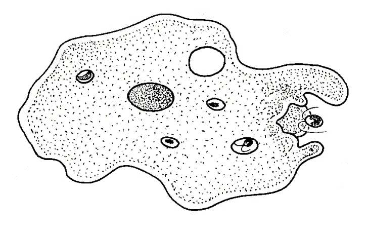
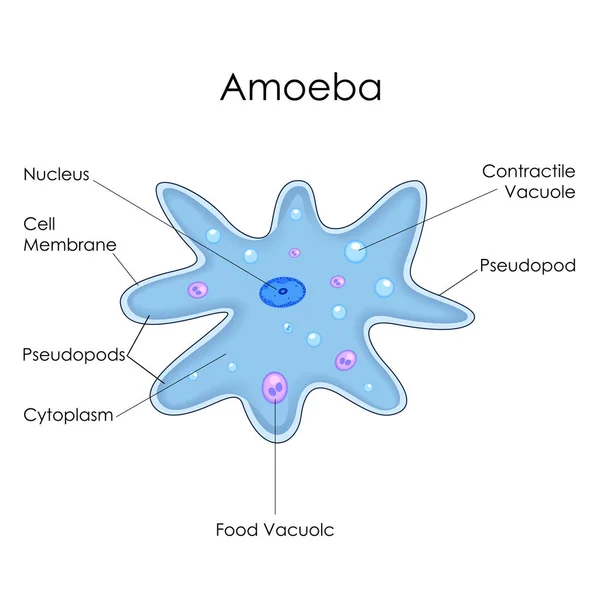
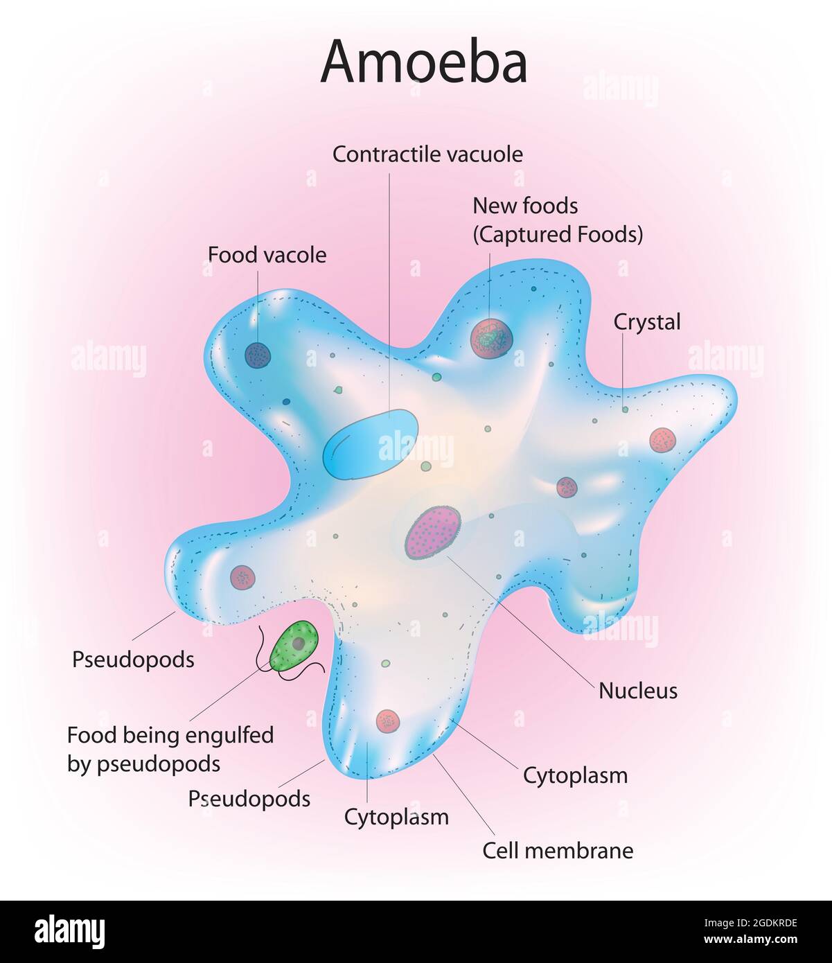
Amoeba diagram labeled. Amoeba proteus is a one-celled animal about 25 mm ( 1/100 inch) in diameter, and is, therefore, invisible to the naked eye. Under the compound microscope it appears as an irregular colorless particle of animated jelly which is constantly changing its shape by thrusting out finger-like processes. Habitat of Amoeba proteus. A paramecium is a unicellular (one cell) eukaryotic organism generally found in stagnant water. While very small, sometimes large paramecium can be seen as tiny specks darting around in a water sample. Paramecium can be about 0.5 mm long. Species of Paramecium range in size from 50 to 330 micrometres (0.0020 to 0.0130 in) in length. Label Amoeba Diagram Using the definitions listed below, label the amoeba. More on Amoebas More Label Me Printouts cell membrane - the thin layer of protein and fat that surrounds the amoeba; it allows some substances to pass into the cell, and blocks other substances. 1. Editable Vector .AI file. 2. Editable Vector .EPS-10 file. 3. High-resolution JPG image. Use for everything except reselling item itself. Description: Amoeba labeled vector illustration. Educational single cell animal structure scheme with ectoplasm, nucleus, mitochondrion, pseudopod, lysosome and contractile vacuole.
amoeba answers com, euglena diagrams diagram link, lab 3 euglena 7b science labs, image gallery euglena labeled keywordsuggest org, protist cell diagram labeled 2003 ford expedition wiring, a draw a well labelled diagram of euglena b name the, the structure and life cycle of amoeba with diagram, Click here👆to get an answer to your question ️ Describe the process of nutrition in amoeba.Draw labelled diagram to show that various steps of nutritions in amoeba. Structure of amoeba primarily encompasses 3 parts - the cytoplasm, plasma membrane and the nucleus. The cytoplasm can be differentiated into 2 layers - the outer ectoplasm and the inner endoplasm The plasma membrane is a very thin, double-layered membrane composed of protein and lipid molecules. #amoeba #sciencediagram #adimushowA beautiful drawing of an Amoeba. And it will teach you to draw the Amoeba very easily. Watch the video and please be kind ...
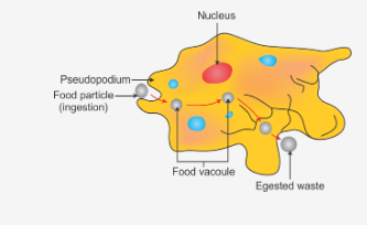
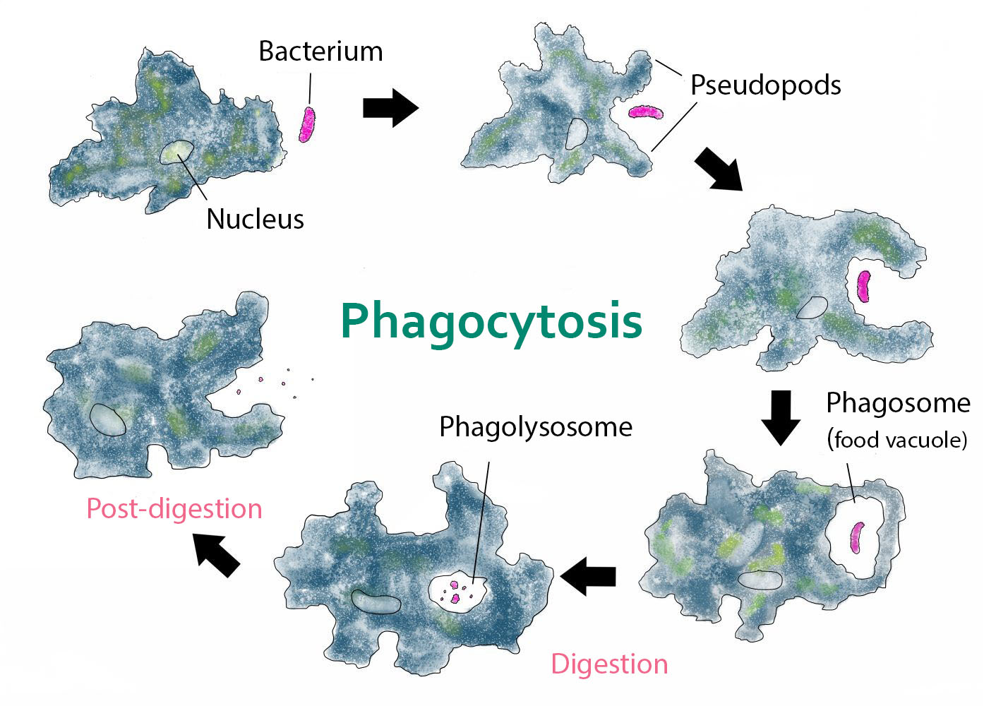
Amoeba Proteus Diagram. Here are a number of highest rated Amoeba Proteus Diagram pictures upon internet. We identified it from obedient source. Its submitted by running in the best field. We take on this nice of Amoeba Proteus Diagram graphic could possibly be the most trending topic next we portion it in google improvement or facebook. Find Labeled Diagram Amoeba Cell stock images in HD and millions of other royalty-free stock photos, illustrations and vectors in the Shutterstock collection. Thousands of new, high-quality pictures added every day. Nutrition in an Amoeba occurs through a process called phagocytosis where the entire organism pretty much engulfs the food it plans on eating up. The mode of nutrition in amoeba is known as holozoic nutrition. It involves the ingestion, digestion and egestion of food material. Amoeba does not have any specialized organ for nutrition. A labelled diagram of Amoeba proteus can be seen above. The pseudopodia are the most defined structures of A. proteus and part of what makes the organism so fascinating. These "false feet" are used for movement and to engulf prey (see Nutrition for further detail) - making it an essential part of its structure.
Solved label the following parts on the diagram below of an ...
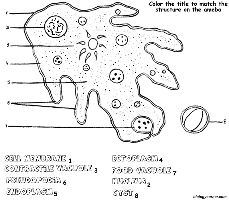
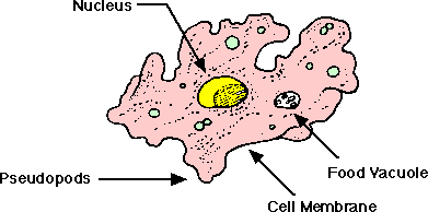
Amoeba (Ameba) Animal Printouts Label Me! Printouts The amoeba is a tiny, one-celled organism. You need a microscope to see most amoebas - the largest are only about 1 mm across. Amoebas live in fresh water (like puddles and ponds ), in salt water, in wet soil, and in animals (including people). There are many different types of amoebas.
Amoeba Diagram Stock Illustrations – 142 Amoeba Diagram Stock ...
Download 110 Amoeba Diagram Stock Illustrations, Vectors & Clipart for FREE or amazingly low rates! New users enjoy 60% OFF. 178,279,237 stock photos online.

Amoeba Diagram Stock Illustrations – 142 Amoeba Diagram Stock ...
An amoeba is unicellular and moves by using pseudopods. A pseudopod is a temporary bulge that forms in the cell membrane as a result of the movement of the cytoplasm. The word pseduopod means "false foot." The pseudopod has two functions, or uses: 1. to move, 2. to capture food.

Amoeba - Mind the Graph
Label diagram of amoeba. Proteus and part of what makes the organism so fascinating. Plural amoebas or amoebae ə ˈ m iː b i often called an amoeboid is a type of cell or unicellular organism which has the ability to alter its shape primarily by extending and retracting pseudopods. An amoeba ə ˈ m iː b ə. It helps the amoeba move feed ...
Draw a neat labelled diagram of Amoeba - CBSE Class 7 Science ...
This will also help you to draw the structure and diagram of amoeba. 1. Fresh water and free living organism commonly available in stagnant water. 2. Body irregular and cytoplasm clearly differentiated into ectoplasm and endoplasm. 3. Body naked, and extends into numerous finger like projections the pseudopodia. 4.

AS Level Biology (9700) P3 Guide – Diagrams – Stude Mate
Download a free printable outline of this video and draw along with us: https://artforall.me/video/how-to-draw-amoeba Thank you for watching. Please subscri...

Lab 1 - Amoebas - 7B Science Labs
Click here 👆 to get an answer to your question ️ Draw Neat Labelled Diagram Of Amoeba shahinakhan7759 shahinakhan7759 18.04.2020 Biology Secondary School answered Draw Neat Labelled Diagram Of Amoeba 2 See answers Advertisement Advertisement abi627780 abi627780
Amoeba anatomy Images, Stock Photos & Vectors | Shutterstock
Amoeba labeled vector illustration. Educational single cell animal structure scheme with ectoplasm, nucleus, mitochondrion, pseudopod, lysosome and contractile vacuole. amoeba structure vector amoeba structure with description Binary fission in amoeba. Vector educational illustration Macrophage Images of macrophages. vector unicellulars set

Amoeba & chlorella
Ameba Coloring

Amoeba Cell Characteristics, Structure, Movement, Nutrition ...

63 Amoeba Diagram Illustrations & Clip Art - iStock
Biological drawings. Amoeba Feeding. Biology teaching ...
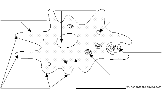
Label Amoeba - EnchantedLearning.com

63 Amoeba Diagram Illustrations & Clip Art - iStock

68 Amoeba diagram Vector Images, Amoeba diagram Illustrations ...
Amoeba anatomy Images, Stock Photos & Vectors | Shutterstock

micro amoeba proteus Diagram | Quizlet
Ameba Coloring
Amoeba - Teaching resources
Amoeba (Ameba) Printout - EnchantedLearning.com
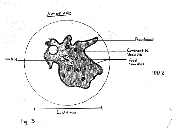
Sample Descriptive Lab Report
Amoeba - Teaching resources
68 Amoeba diagram Vector Images, Amoeba diagram Illustrations ...
Expain amoeba diagram? | EduRev Class 8 Question

Introduction to Protista: Amoeba | Carolina.com
Amoeba - Simple English Wikipedia, the free encyclopedia
Free Ameba Diagram, Download Free Ameba Diagram png images ...
Nutrition in Amoeba - Process, Diagram, Questions | ProtonsTalk
amoeba/amoeba diagram/amoeba image/labelled diagram amoeba/amoeba labelled diagram / science diagram

63 Amoeba Diagram Illustrations & Clip Art - iStock

Sketch and label the diagram of Amoeba and describe it brief ...
Eukaryotic Cell Structure - Sciencetopia

draw a diagram to show nutrition in amoeba and label the ...
Ameba Diagram Labeled - Ameba Shape Clip Art - Free ...

Amoeba Diagram Stock Illustrations – 142 Amoeba Diagram Stock ...

Amoeba PowerPoint Diagram - PSlides | Powerpoint, Diagram ...

Free Ameba Diagram, Download Free Ameba Diagram png images ...

One-Cell Wonder Number One | Sunesis Science
Amoeba - Labeled anatony of amoeba proteus, Detailed vector ...

Life cycle of an amoeba (scheme) and cyst structure. a ...
Draw a neat labelled diagram Amoeba. - Sarthaks eConnect ...


Comments
Post a Comment