43 mitochondria labeled diagram
Week 13B Explore: The Mighty Mitochondria Diagram As the video plays, (remember: you can pause the video at any time) label everything you see labeled in the video on your own diagram of a mitochondria and a phospholipid bilayer. Note: the narrator uses a variety of colors, which is a good idea to use, especially if you are a visual learner or a color learner. › proteins › proteinProtein Targeting (With Diagram) | Molecular Biology ADVERTISEMENTS: Let us make an in-depth study of the protein targeting. After reading this article you will learn about: 1. Introduction to Protein Targeting 2. Signal Sequence 3. Transport of Proteins into ER 4. Signal Sequence Recognition Mechanism 5. Role of Golgi Complex in Protein Transportation 6. Transport of Proteins from Golgi to Lysosomes 7. […]
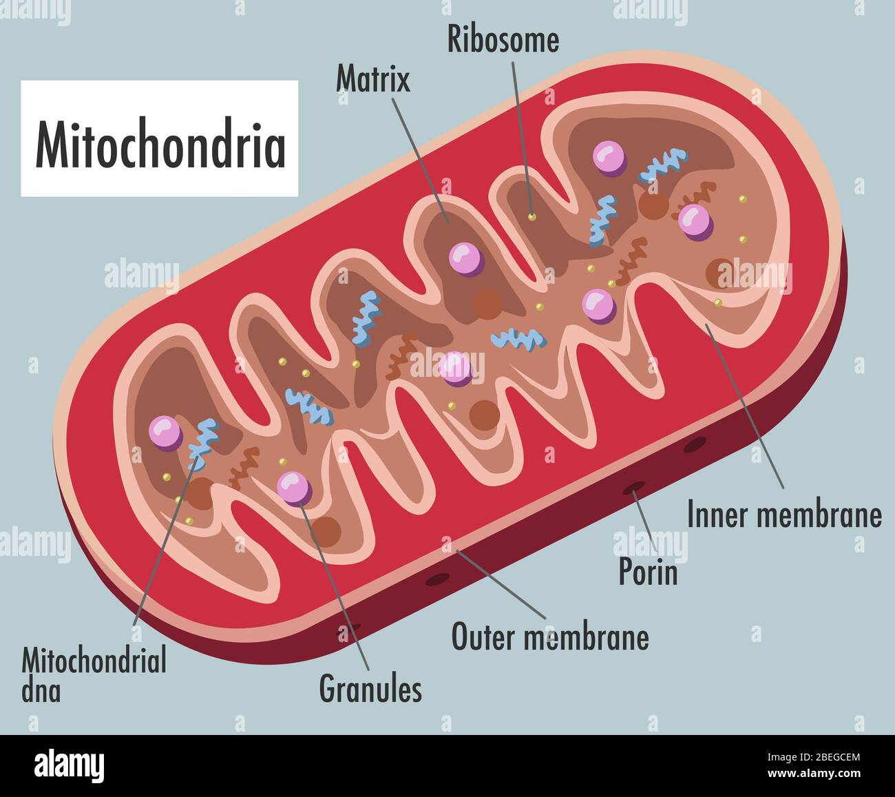
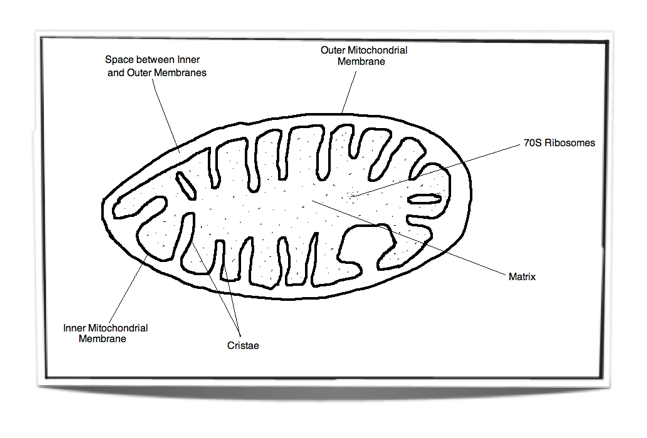
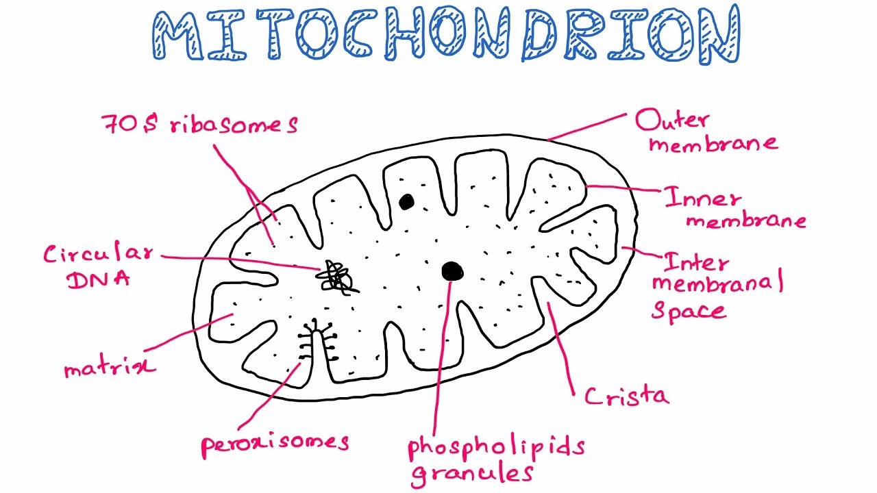
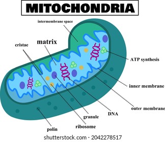
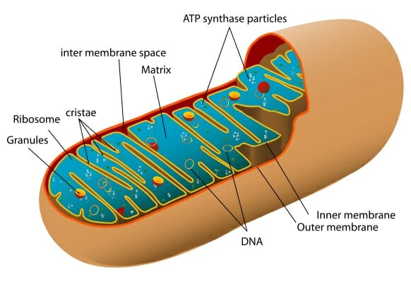
Cell Organelles- Definition, Structure, Functions, Diagram Structure of Mitochondria. A mitochondrion contains two membranes with the outer layer being smooth while the inner layer is marked with folding and finger-like structures called cristae. The inner mitochondrial membrane contains various enzymes, coenzymes, and components of multiple cycles along with pores for the transport of substrates, ATP ...
Mitochondria labeled diagram
Plant Cell Diagram Mitochondria Labeled : Functions and ... Plant Cell Diagram Mitochondria. This basic structure of a plant cell is shown below - the same plant cell, as viewed with the light microscope, and with the transmission electron microscope. It is easier to describe these parts by using diagrams: Animal cells and plant cells also contain tiny objects called mitochondria in their cytoplasm. Mitochondria Quiz - PurposeGames.com Mitochondria are the "power houses" of cells. Eukaryote cells, meaning animal cells (more advanced organisms) are the type of cells that have them, and they need mitochondria to function properly. This quiz has tags. Click on the tags below to find other quizzes on the same subject. Label the Structures of the Mitochondria Quiz Label the internal and external structure of mitochondria . This game is part of a tournament. You need to be a group member to play the tournament
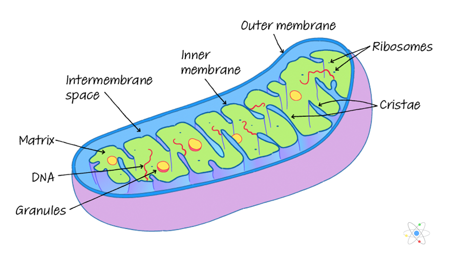
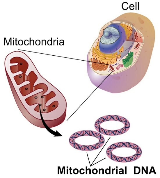
Mitochondria labeled diagram. | Intercellular mitochondrial transfer between astrocytes ... Download scientific diagram | | Intercellular mitochondrial transfer between astrocytes. (A) Schematic showing that human astrocytes (HA) were labeled with GFP or mitodsred and then co-cultured at ... Oxidative Phosphorylation of Mitochondrion (With Diagram) Oxidative Phosphorylation of Mitochondrion (With Diagram) Linkage of phosphorylation to oxidative metabolism was first proposed in the 1930s. The first evidence was the observation that phosphate was removed from the medium when TCA cycle intermediates were metabolized by cells. Subsequently, it was shown that the disappearance of inorganic ... Structure of Mitochondria | Biology diagrams, Cell biology ... Oct 12, 2015 - The structure of mitochondria is essential knowledge for students of cell biology. Mitochondria are cell organelles whose overall shape resembles rounded rods and is often drawn in 2D as an oval-shape. Mitochondria have a double-membrane structure and contain many substructures including enzymes, ribosomes and mitochondrial DNA (mtDNA). Diagram of Mitochondria | Labelled Diagram of Mitochondria A well-labelled diagram of mitochondria is given below for your better understanding of the structure. Labelled Diagram of a Mitochondrion Image will be updates soon Characteristics of the Mitochondrial DNA/ Genome: The mitochondrial DNA is circular and is made up of 16,569 DNA base pairs.
The diagram is schematic representation of the electron ... The diagram is schematic representation of the electron transport mechanism in mitochondria. Label the diagram to complete the model. 2 See answers Advertisement Advertisement river2738 river2738 The big blue area is the mitochondrial matrix. This is where the citric acid cycle takes place; also known as the Krebs cycle or Tricarboxylic acid cycle. Mitochondrial Function - Biology Wise This BiologyWise article the structure and function of mitochondria with the help of a labeled diagram. The study of cell organelles and their functions covers individual responsibilities of each subunit of a cell - each organelle is a structure within a cell that has a specific function. Fluorescence imaging of mitochondria and cytoskeleton. (A ... Download scientific diagram | Fluorescence imaging of mitochondria and cytoskeleton. (A) F-actin cytoskeleton labeled with A488-PHD. (B) Mitochondria labeled with DsRed2. (C) Overlay of (A) and (B ... Mitochondria Labeling Diagram | Quizlet The electron transport chain and chemiosmosis takes place on this membrane as part of cellular respiration to create ATP. The cristae increase the surface area of the inner membrane, allowing for faster production of ATP because there are more places to perform the process.
PDF Mitochondria, Their Structure, Functions, and Diseases ... The fusion fission cycle of mitochondria. C). Internal structure. D). MtDNA, structure and packaging. ... labeled mitochondria showing the looping of the reticulum throughout its length. transitioning of mitochondria between punctate and reticulum states through alternating fission and fusion is now known to be critical to Difference Between Mitochondria and Chloroplast: Parts ... Given below is a well- labeled diagram of a mitochondria to help in better understanding of its structure. Structure of Mitochondria. What are Chloroplasts? Besides plant cells, chloroplasts are also found in algae. They are located in the cytosol of a cell, and contain chlorophyll to absorb solar energy. › fungi › structure-ofStructure of Fungal Cell (With Diagram) | Fungi The mitochondria function as the power house of the cell. There is no fundamental difference between the mitochondria of fungi and those of green plants. However, Hawker (1965) holds that the cristae of fungal mitochondria are fewer, flatter and more irregular than those of the green plants. (iii) Golgi Apparatus (Dictyosomes): Animal Cell Diagram Mitochondria Labeled : Functions and ... Animal Cell Diagram Mitochondria Labeled. Tuesday, April 27th 2021. | Diagram. Animal Cell Diagram Mitochondria. Animal cells have a basic structure. The mitochondrion (plural mitochondria) is a membrane-bound organelle found in the cytoplasm of eukaryotic cells.
Mitochondria, Illustration, Labeled Stock Photo - Alamy
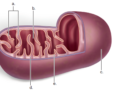
Glycolysis Worksheet Diagram - schematron.org Glycolysis Worksheet Diagram. Use the words below to label the diagram of cellular respiration on the lines provided. Glycolysis uses ATP to break a molecule of glucose in half, pro-. Explain, in general terms, how carbohydrates are oxidized by glycolysis and Krebs cycle to Practice: Mitochondria Structure Label workbook diagram 1.

With a labelled diagram, explain the structure and function ...
Structure of Mitochondria (With Diagram) | Botany This will also help you to draw the structure and diagram of mitochondria. 1. Mitochondria are commonly called the "Power house" of the cell. 2. Benda (1897) was the first to coin the term mitochondrion. 3. Usually, mitochondria are 0.5 to 1 n in diameter and 3-6n in length.
Mitochondrion & Cellular Respiration Diagram Worksheet
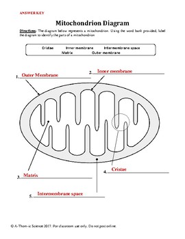
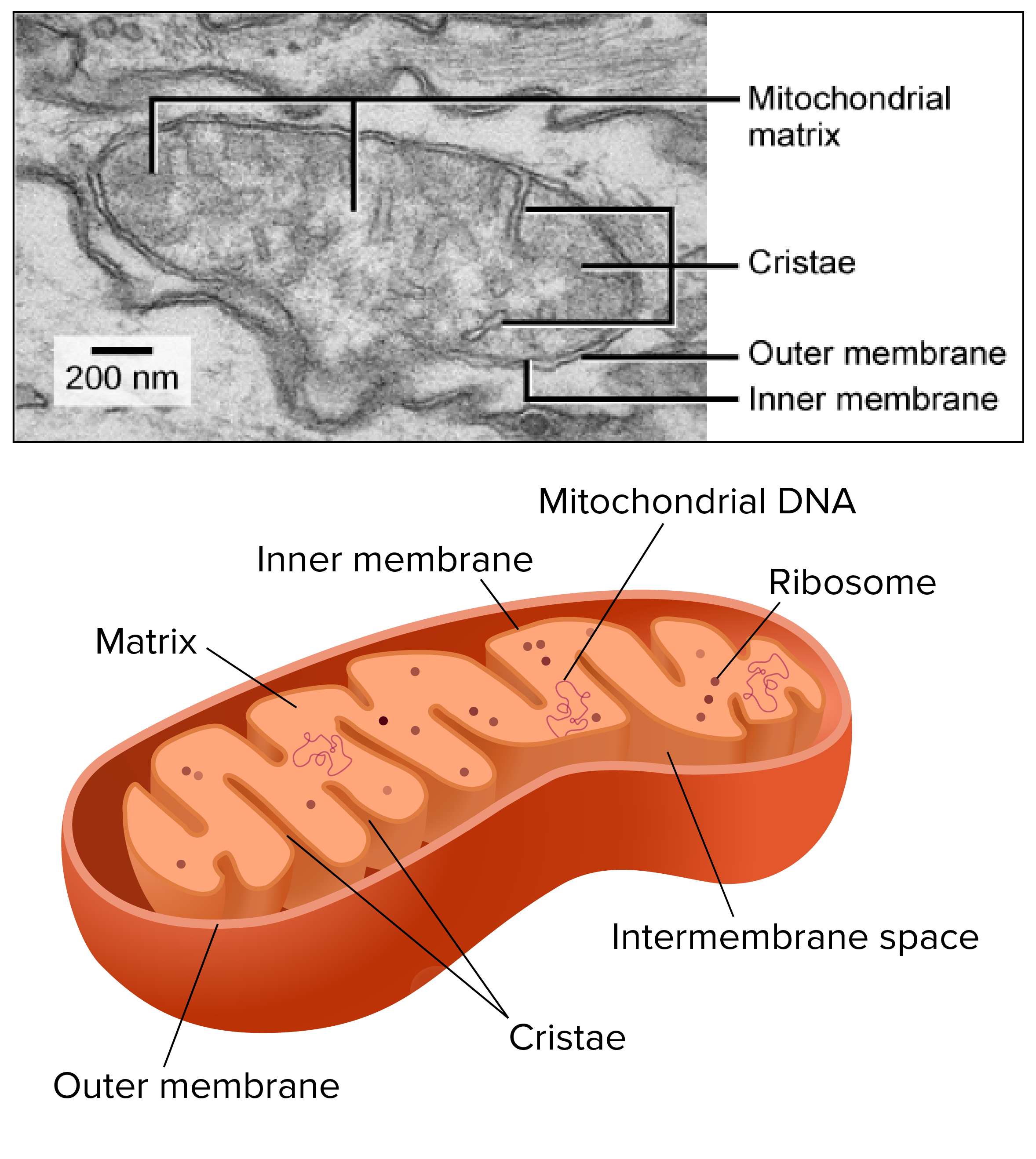
Diagram of a mitochondrion — Science Learning Hub Diagram of a mitochondrion. ADD TO COLLECTION. Add to new collection. CANCEL. Tweet. Rights: University of Waikato Published 20 July 2009 Size: 28 KB Referencing Hub media. Diagram of a mitochondrion showing the inner and outer membranes, and the folded cristae.
Short answer question:Draw a well-labelled sketch of the ...
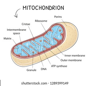
What Is Mitochondria (Structure, Diagram & Function) Mitochondria diagram explaining the structure of mitochondria Structure of Mitochondria The mitochondrion is a double-membraned, rod-shaped structure found in both plant and animal cell. Its size ranges from 0.5 to 1.0 micrometre in diameter. The structure comprises an outer membrane, an inner membrane, and a gel-like material called the matrix.

Diagram of a Mitochondrion
Mitochondria Labeling Diagram | Quizlet Start studying Mitochondria Labeling. Learn vocabulary, terms, and more with flashcards, games, and other study tools.
How Does the Mitochondria Produce Energy for the Cell
A Labelled Diagram Of Mitochondria with Detailed Explanation Mitochondria are a double-membrane-bound cell organelle found in most eukaryotic organisms. In all living cells, these cell organelles are found freely floating within the cytoplasm of the cell. The diagram of Mitochondria is useful for both Class 10 and 12.
IB Biology Notes - 8.1 Cell respiration
draw a well labelled diagram indicating the synthesis of ... What is beta-oxidation of fat(in mitochondria)? Please Explain the points 'b' & 'c' of chemiosmotic hypothesis of photosynthesis with the help of a diagram Where does the electron transport system take place in the mitochondrion? Name the component which transfers electrons from ubiquinol to cytochrome c.

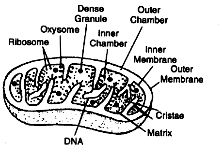
Parts of a Mitochondria Diagram | Ribosomes & Function of ...
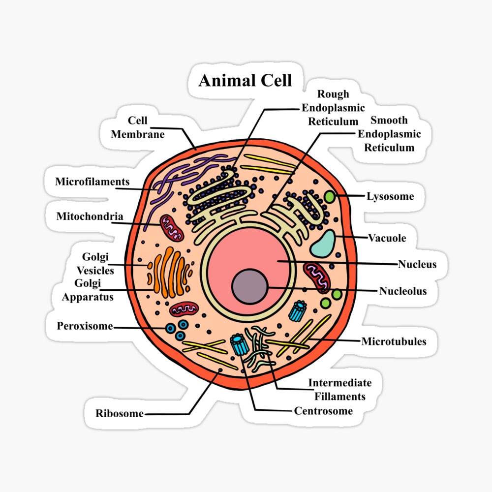
Animal Cell Diagram Labeled | EdrawMax Template As you can see from the following labeled diagram, animal cells are eukaryotic cells that contain a membrane-bound nucleus. According to the animal cell labeled diagram, some of the cell organelles of an animal cell are the cell membrane, Cytosol, Cytoskeleton, nucleus, Ribosomes, Endoplasmic Membrane, Vesicles, Mitochondria, and more. Creating ...
![Solved] Draw a labelled diagram of mitochondria write the ...](https://srv-supersonic-images.z-dn.net/editor_images/f2597698-8ed1-4f78-82d0-ef96ce864d3a.png)
Solved] Draw a labelled diagram of mitochondria write the ...
The diagram is schematic representation of the electron ... The diagram is schematic representation of the electron transport mechanism in mitochondria. Label the diagram to complete the model. 2 See answers Advertisement smilodon As shown in the presented diagram in the process of electron transport happens in the mitochondrial matrix.

How to monitor mitophagy in mammalian cells | tebu-bio's blog
Label the Structures of the Mitochondria Quiz Label the internal and external structure of mitochondria . This game is part of a tournament. You need to be a group member to play the tournament

Mitochondrion & Cellular Respiration Diagram Worksheet ...
Mitochondria Quiz - PurposeGames.com Mitochondria are the "power houses" of cells. Eukaryote cells, meaning animal cells (more advanced organisms) are the type of cells that have them, and they need mitochondria to function properly. This quiz has tags. Click on the tags below to find other quizzes on the same subject.

Structure of Mitochondria | Biology diagrams, Cell biology ...
Plant Cell Diagram Mitochondria Labeled : Functions and ... Plant Cell Diagram Mitochondria. This basic structure of a plant cell is shown below - the same plant cell, as viewed with the light microscope, and with the transmission electron microscope. It is easier to describe these parts by using diagrams: Animal cells and plant cells also contain tiny objects called mitochondria in their cytoplasm.
Mitochondria: Definition, Structure & Function (with Diagram)

Mitochondria Functions, Location, Diagram and Structure ...
![Solved] Draw a labelled diagram of mitochondria write the ...](https://hi-static.z-dn.net/files/dc5/293ca3f41fbb858652c55b5f6210947f.jpg)
Solved] Draw a labelled diagram of mitochondria write the ...
Mitochondrion: Definition, Structure and Function | Biology ...

Mitochondria Diagram Stock Illustrations – 560 Mitochondria ...
Labeled Animal Cell Diagram" Photographic Print by BundaBear ...

435 Mitochondria Vector Images, Mitochondria Illustrations ...

Mitochondria Labeling Lentivirus (MTS)

The Cytoplasm and Cellular Organelles | Anatomy and Physiology I

Schematic diagram showing different processes of mitochondria ...

Muscle membrane vector illustration. Labeled scheme with myofibril, disc, zone, line and band. Anatomical diagram with mitochondria, sarcoplasm, ...

Purification of SILAC-labeled mitochondria from SY5Y cells. A ...
how to draw the mitochondria
Mitochondria Images, Stock Photos & Vectors | Shutterstock
Mitochondria Handout
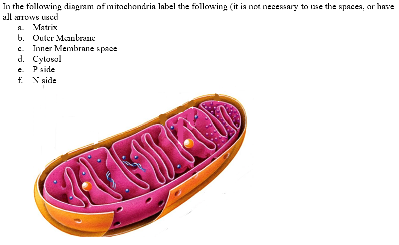
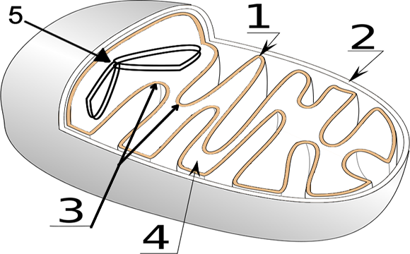
Solved In the following diagram of mitochondria label the ...
Inner mitochondrial membrane Images, Stock Photos & Vectors ...

Mitochondria Functions, Location, Diagram and Structure ...

1,860 Mitochondria Stock Photos and Images - 123RF

Mitochondria | BioNinja

Mitochondria: Structure, Functions and Diagram – StudiousGuy
4 Draw the labeled diagram of mitochondria - Science - The ...
Mitochondria - Definition, Function & Structure | Biology ...
Draw a neat and labelled diagram of mitochondria. - Sarthaks ...

How to draw mitochondria | Diagram | Easy and well labelled diagram |NCERT | Power house of the cell
Solved: Label this diagram of a mitochondrion, and state a ...

Mitochondrion Clip Art - Royalty Free - GoGraph
Diagram of Mitochondria and Function of Mitochondria Parts ...
Mitochondria and chloroplasts (article) | Khan Academy
INTRODUCTION

How to draw the diagram of mitochondria //easy steps by step ...


Comments
Post a Comment