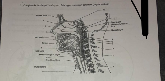
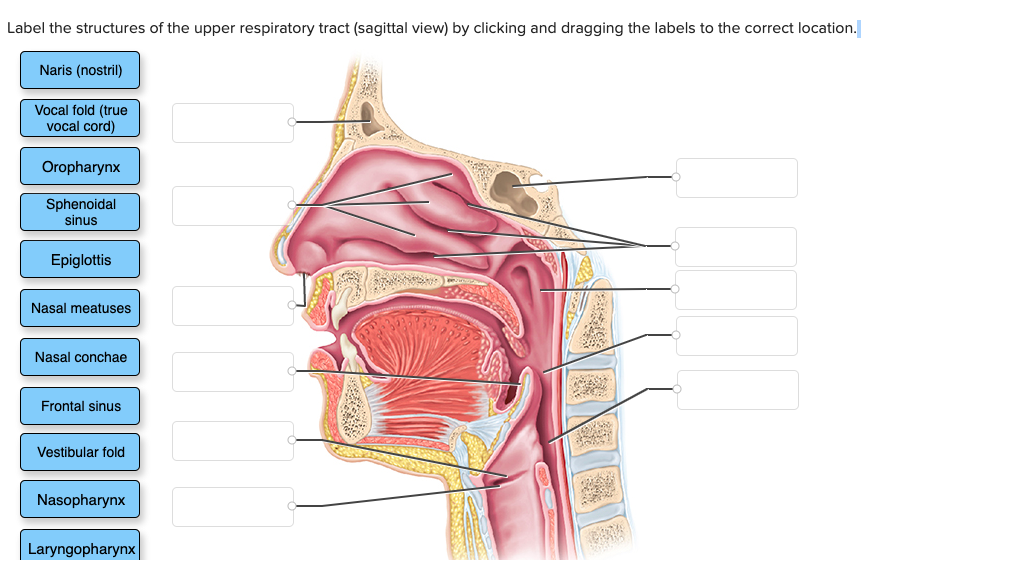
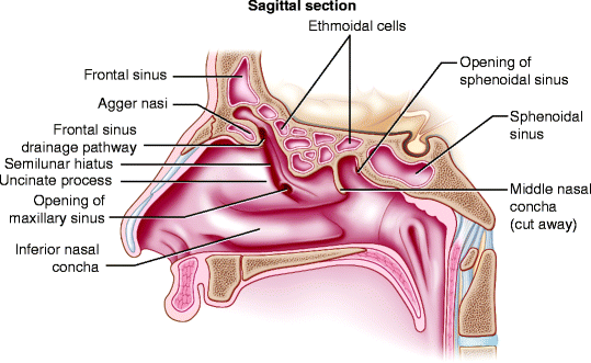
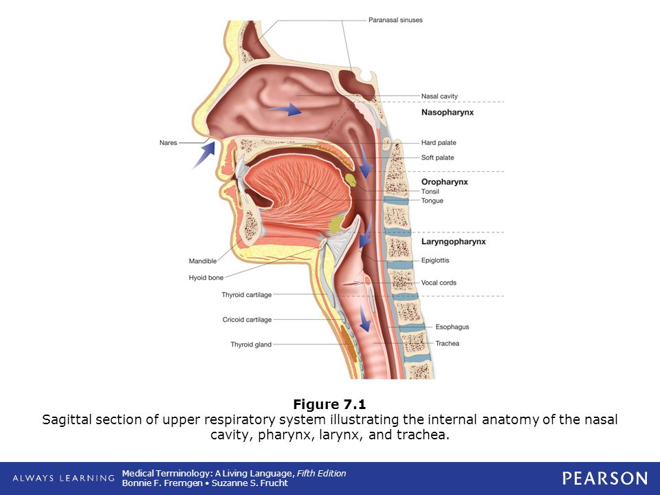
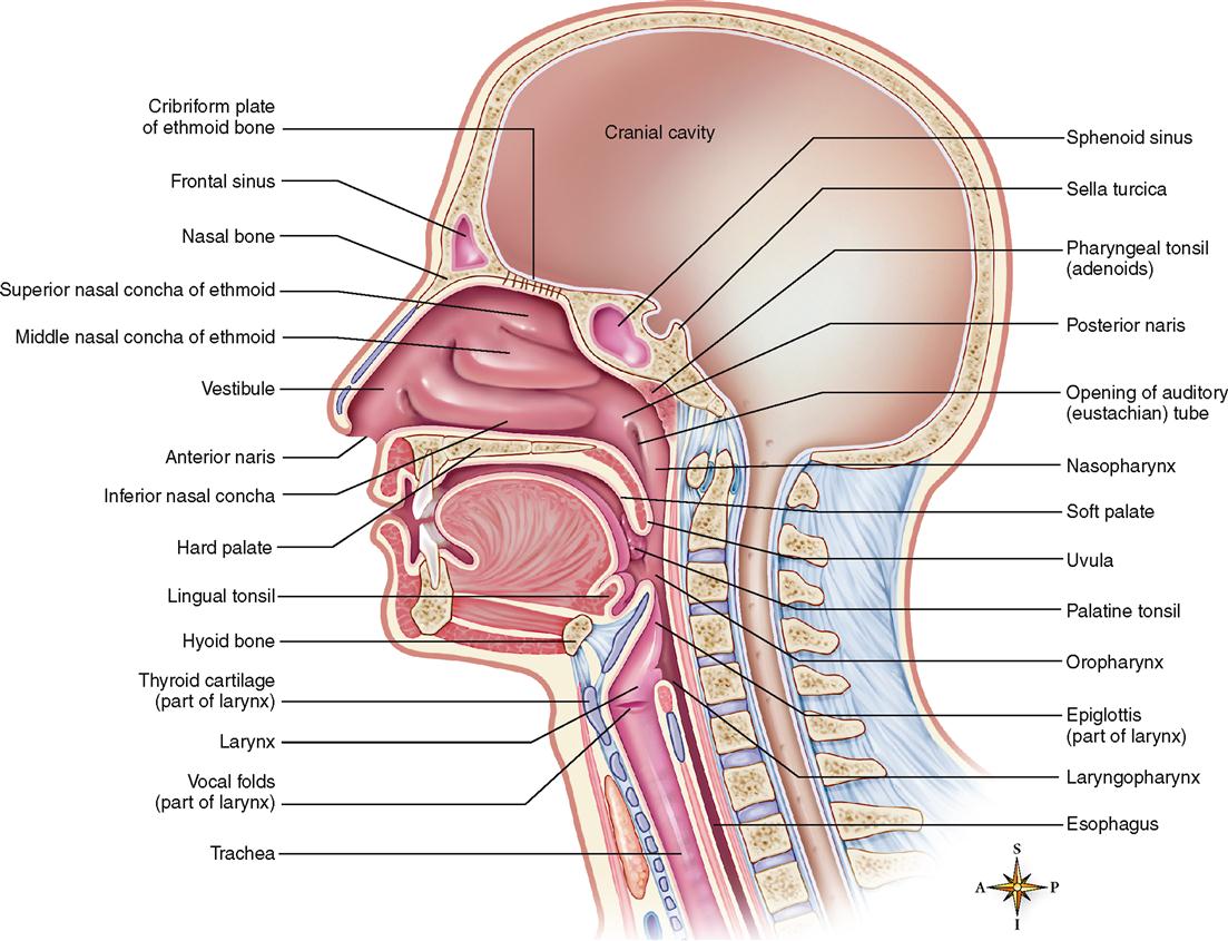
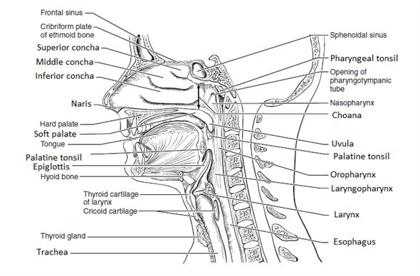
38 complete the labeling of the diagram of the upper respiratory structures (sagittal section)
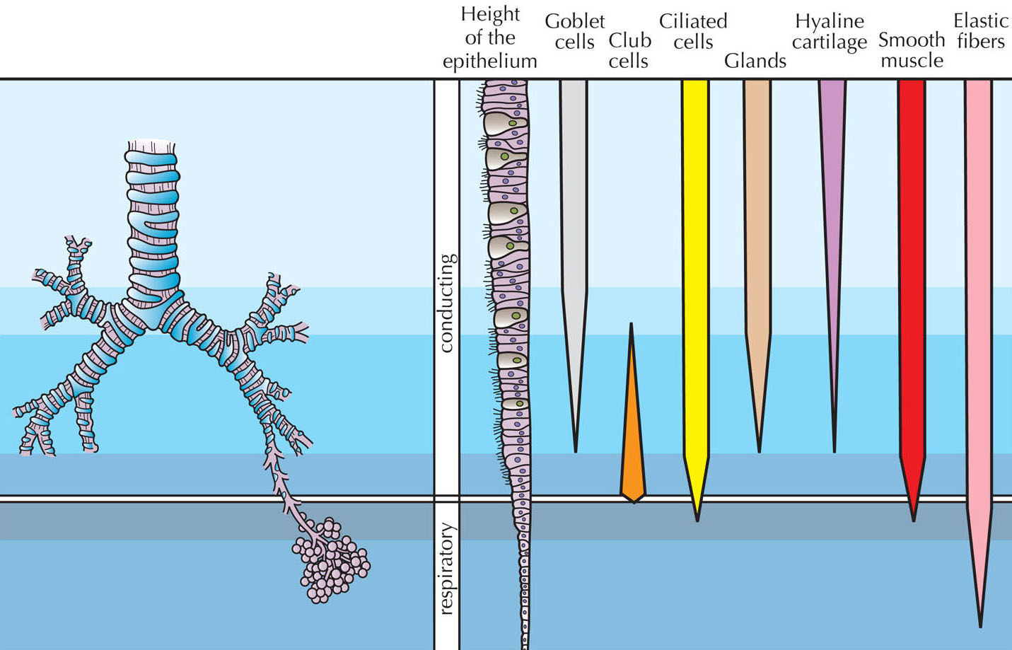
The structures that form these walls vary depending on which segment of the ventricle is being viewed. Let's walk through the boundaries of the anterior horn, body, posterior horn, and inferior horn. We will also look at sagittal views and coronal views of the brain/ventricles to help illustrate the boundaries. Anterior (Frontal) Horn The division of the respiratory system into conducting and respiratory airways delineates their function and roles. The conducting portion, consisting of the nose, pharynx, larynx, trachea, bronchi, and bronchioles, which all serve to humidify, warm, filter air. The respiratory portion is involved in gas exchange. There are three major types of ...
The mediastinum is an area found in the midline of the thoracic cavity, that is surrounded by the left and right pleural sacs.It is divided into the superior and inferior mediastinum, of which the latter is larger.. The inferior mediastinum is further divided into the anterior, middle and posterior mediastinum.Every compartment of the mediastinum contains many vital organs, vascular and neural ...
Complete the labeling of the diagram of the upper respiratory structures (sagittal section)
Upper and Lower Respiratory System Structures 1. Complete the labeling of the diagram of the upper respiratory structures (sagittal section). Rating: 4.7 · 12 votes The thorax is the region between the abdomen inferiorly and the root of the neck superiorly.[1][2] It forms from the thoracic wall, its superficial structures (breast, muscles, and skin) and the thoracic cavity. Upper and Lower Respiratory System Structures. 1. Complete the labeling of the diagram of the upper respiratory structures (sagittal section).4 pages
Complete the labeling of the diagram of the upper respiratory structures (sagittal section). Upper and lower Respiratory System Structures. 1. Complete the labeling of the diagram of the upper respiratory structures (sagittal section).4 pages Spinal Cord Cross Section. Looking at a cross section of the spinal cord, you would see gray matter shaped like a butterfly surrounded by white matter. The gray matter is the core and ends up to be four projections that are known as horns. At the back are two dorsal horns and away from the back are two ventral horns. The larynx is a cartilaginous segment of the respiratory tract located in the anterior aspect of the neck. The primary function of the larynx in humans and other vertebrates is to protect the lower respiratory tract from aspirating food into the trachea while breathing. It also contains the vocal cords and functions as a voice box for producing sounds, i.e., phonation. Function and anatomy of the heart made easy using labeled diagrams of cardiac structures and blood flow through the atria, ventricles, valves, aorta, pulmonary arteries veins, superior inferior vena cava, and chambers. Includes an exercise, review worksheet, quiz, and model drawing of an anterior vi
NAM LAB TIME/DATE_ Anatomy of the Respiratory System Upper and Lower Respiratory ... of the diagram of the upper respiratory structures (sagittal section). The last chapter of this human anatomy module presents anatomical sections of the lower limb, focusing on the gluteal region, the thigh, the femoral region, a section of the popliteal fossa, anatomical sections of the leg, an axial section of the ankle, a frontal section of the tarsus area and a frontal section of the forefoot. Anatomy and Physiology Workbook. One of the best Anatomy and Physiology Coloring Book believe it or not Workbook. 7Anatomy and physiology coloring workbook ch 7 A complete study guide 12th edition 9780134459363. 18 Anatomy And Physiology Coloring Workbook Chapter12 Answers anatomy and physiology coloring workbook answer key. 15The Anatomy Physiology Coloring Workbook has been created ... A&P II - Review Sheet 36 - Anatomy of the Respiratory System ... Image: Know and be able to label the following. Two pairs of vocal folds are found in the ... Rating: 5 · 4 reviews
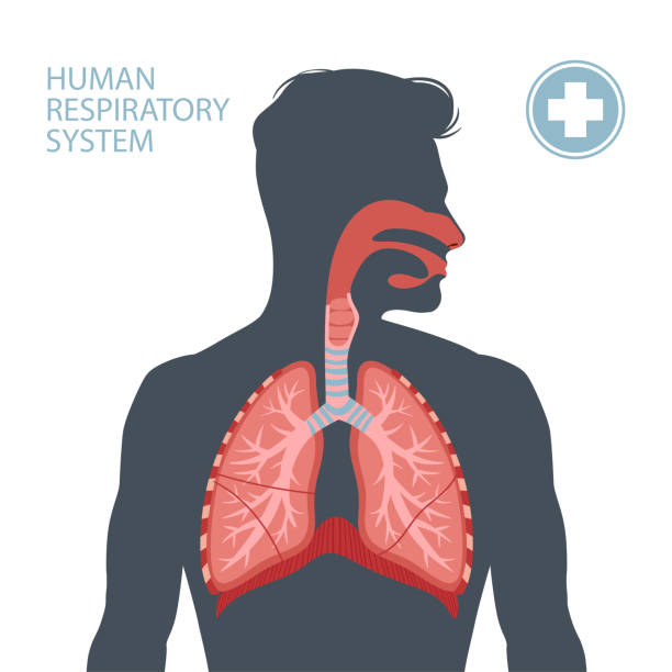
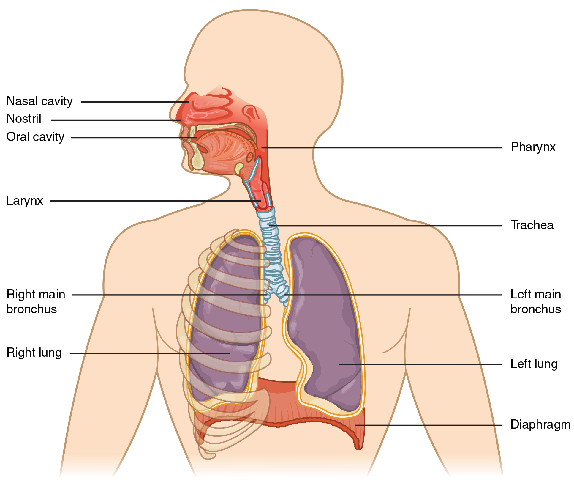
EXERCISE 36 REVIEW SHEET Anatomy of the Respiratory System Name Lab Time/Date pper and Lower Respiratory System Structures the diagram of the upper respiratory structures (sagittal section) Opening of pharyngotympanic Hard palate Hyoid bone Thyroid... In this section, let us get into some more detailed study of a few of the essential structures of the lung, which can be appreciated better under an electron microscope. Respiratory Bronchiole: This is the region of transition between conducting and respiratory portions (where the exchange of gases begins). The respiratory tract has two major divisions: the upper respiratory tract and the lower respiratory tract. The organs in each division are shown in Figure 16.2. 2. In addition to these organs, certain muscles of the thorax (the body cavity that fills the chest) are also involved in respiration by enabling breathing. The main function of the trachea is to transport air from the upper respiratory tract to the lungs. Air that enters the trachea is warmed and moisturized before moving on to the lungs. Mucus on the trachea walls can catch debris or particles. This debris is then transported upward by cilia, tiny hair-like structures that remove it from the airway.

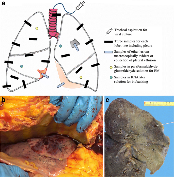
Pulmonary Pathology And Covid 19 Lessons From Autopsy The Experience Of European Pulmonary Pathologists Springerlink
Full labeled anatomical diagrams - Anatomy of the abdomen and digestive system: these general diagrams show the digestive system, with the major human anatomical structures labeled (mouth, tongue, oral cavity, teeth, buccal glands, throat, pharynx, oesophagus, stomach, small intestine, large intestine, liver, gall bladder and pancreas).
The femur is the primary bone of the leg. It supports the weight of the body on the leg and is capable of carrying 30 times the weight of the body. The femur provides the ability for articulation and leverage for the leg. Articulation allows for standing, walking, and running. The femur is the primary bone of the leg and all other leg bones are ...
MRI of the upper extremity anatomy - Atlas of the human body using cross-sectional imaging. We created an anatomical atlas of the upper limb, an interactive tool for studying the conventional anatomy of the shoulder, arm, forearm, wrist and hand based on an axial magnetic resonance of the entire upper limb. Anatomical structures and specific ...

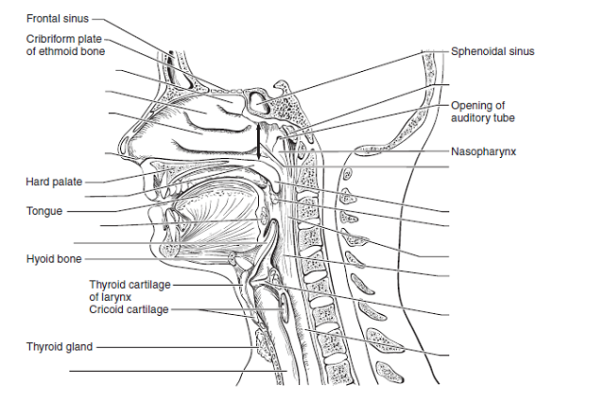
Part A The Respiratory System Drag Each Label To The Appropriate Location On This Diagram Homeworklib
Coronal section of the kidney. The kidneys are a pair of bean-shaped organs located on either side of the superior posterior abdominal wall. Its lateral border is convex while its medial border is concave. The medial concavity is the point at which the renal neurovascular structures enter and leave the kidneys.
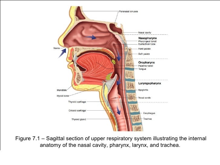
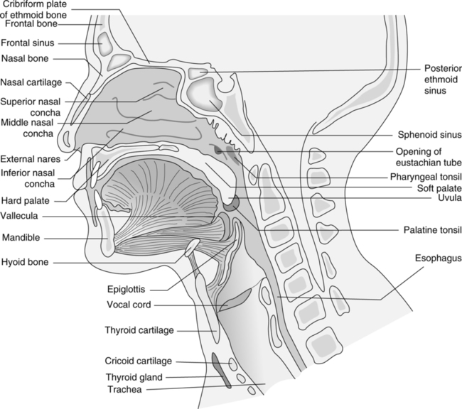
1. Complete the labeling of the diagram of the upper respiratory structures (sagittal section). ... Epiglottis -----f--;f. ... 2. Two pairs of vocal folds are found ...
The module on the anatomy of the brain based on MRI with axial slices was redesigned, having received multiple requests from users for coronal and sagittal slices. The elaboration of this new module, its labeling of more than 524 structures on 379 MRI images in three different views and on 26 anatomical diagrams, took more than 6 months.

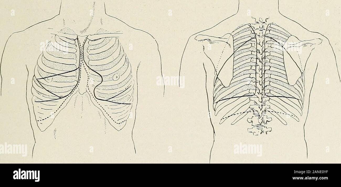
Figure 22 3 The Upper Respiratory Tract Midsagittal Section Of The Head And Neck Medical Anatomy Joints Anatomy Anatomy And Physiology
Skull The skull is a strong, bony capsule that rests on the neck and encloses the brain. It consists of two major parts: the neurocranium (cranial vault) and the viscerocranium (facial skeleton). The neurocranium is the part enveloping the brain and is formed out of two parts; the skull base that supports the brain and the calvaria (skullcap) that sits on top of the base, covering the brain.
quizzes, and labeling diagrams. Suggestions are made for group projects and practical tasks. Each chapter contains facts and key points in bullet-point format for quick reference, followed by several different activities designed to aid memory and support study. Fun and interactive exercises make this core subject
2 figure 5415 label the features associated with the liver and pancreas. Start studying cadaver with pictures. Label the structures of the spleen. Figure 5414 label the features of the stomach and nearby regions in this frontal section of a cadaver anterior view. Label the internal features of stomach and duodenum using the hints if provided.
39 Parts Of Throat Diagram. Weld Root: As you can see in the diagram of a weld above, the root of a weld is where the bottom or underside of a weld crosses the surface of the base metal. Fillet Weld Throat: When you discuss the throat of a weld there are two to consider: 1) theoretical weld throat 2) actual weld throat.
Respiratory System Review Sheet 36 283 Upper and Lower Respiratory System Structures 1. It includes the nose mouth larynx trachea bronchial tubes lungs diaphragm and muscles that enable breathing. Complete the labeling of the diagram of the upper respiratory structures sagittal section.
Upper digestive tract (sagittal view) The pharynx, more commonly known as the throat, is a five cm long tube extending behind the nasal and oral cavities until the voice box and the esophagus.Essentially, it forms a continuous muscular passage for air, food, and liquids to travel down from your nose and mouth to your lungs and stomach.. The functions of the pharynx are accomplished by two sets ...
Complete the labeling of the diagram of the upper respiratory structures (sagittal section). 2. Two pairs of vocal folds are found in the larynx. Rating: 5 · 2 reviews
Upper and Lower Respiratory System Structures 1. Complete the labeling of the diagram of the upper respiratory structures (sagittal section). Rating: 4.7 · 12 votes
Cross-sectional labeled anatomy of the head and neck of the domestic cat on CT imaging (bones of the skull, cervical spine, mandible, hyoid bone, muscles of the neck, nasal cavity and paranasal sinuses, oral cavity, larynx)
The skull lateral view is a non-angled lateral radiograph of the skull. This view provides an overview of the entire skull rather than attempting to highlight any one region. Indications This projection is used to evaluate for skull fractures, ...
Coronal section of the brain at the level of the thalamus. The frontal and temporal lobes are observed in their previously described locations. The body of the corpus callosum forms the roof of the body of the left and right lateral ventricles, which are separated from each other by the septum pellucidum.The insula of Reil and the Sylvian fissure maintain their lateral relationship to the ...

Complete The Labeling Of The Diagram Of The Upper Respiratory Structures Sagittal Section Course Hero
Respiratory System SHEET Upper and Lower Respiratory System Structures 1. Complete the labeling of the model of the respiratory structures (sagittal section) shown below. Nasal "Conchal Nasal meatus REVIEW as a distibule Hard plate -posterior nasal aperature -s of t palate "uvula -palentine tonsil epiglottis vestibular fold vocal fold Thyroid ...

Complete The Labeling Of The Diagram Of The Upper Respiratory Structures Sagittal Section Course Hero
The inside of the nose, including the bones, cartilage and other tissue, blood vessels and nerves, all the way back posteriorly to the nasopharynx, is called the nasal cavity. It is considered part of the upper respiratory tract due to its involvement in both inspiration and exhalation.
Upper and Lower Respiratory System Structures 1. Complete the labeling of the diagram of the upper respiratory structures (sagittal section). Rating: 4.7 · 12 votes
Upper and Lower Respiratory System Structures. 1. Complete the labeling of the diagram of the upper respiratory structures (sagittal section).4 pages
The thorax is the region between the abdomen inferiorly and the root of the neck superiorly.[1][2] It forms from the thoracic wall, its superficial structures (breast, muscles, and skin) and the thoracic cavity.
Upper and Lower Respiratory System Structures 1. Complete the labeling of the diagram of the upper respiratory structures (sagittal section). Rating: 4.7 · 12 votes

Complete The Labeling Of The Diagram Of The Upper Respiratory Structures Sagittal Section Course Hero











Comments
Post a Comment