41 dissecting microscope diagram
May 11, 2018 — Dissecting microscope parts include separate objective lenses and eyepieces. As a result, you have two separate optical paths for each eye. The ... Dissecting microscopes are used for viewing live specimens or three-dimensional objects too large or thick to be accommodated by compound microscopes. Specimens can be physically manipulated under magnification, since they do not have to be mounted onto a slide for observation under a dissecting microscope. These ...
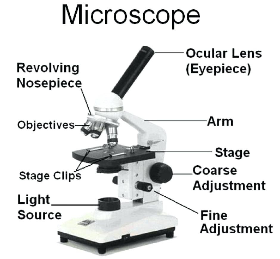
Microscope Parts and Functions With Labeled Diagram and Functions How does a Compound Microscope Work?. Before exploring microscope parts and functions, you should probably understand that the compound light microscope is more complicated than just a microscope with more than one lens.. First, the purpose of a microscope is to magnify a small object or to magnify the fine details of a larger ...
Dissecting microscope diagram
Dissecting microscopes; Monocular, Binocular, and Trinocular microscopes; In addition to our selection of affordable microscopes for sale, we also offer a wealth of free resources. Find worksheets and experiments, a helpful diagram, a gift guide, a brief history of microscopes, and more. A stereo microscope is defined as a type of microscope that provides a three-dimensional view of a specimen. It is also known as a dissecting microscope. In a stereo microscope, there are separate objective lenses and eyepiece such that there are two separate optical paths for each eye. Stereo Microscope Diagram. Principle of Stereo Microscope 5 Important Types Of Microscopes Used In Biology With Diagram. Solved 1 Familiarise Younelf With All The Parts Of The M. Stereo Microscope Basics. Microscope Parts Function Biological Science Picture Directory. Biology 2404. Http Www Lamission Edu Lifesciences Bio3labs Bio3 20lab03 Sp12 Microscopy 20 20cells Pdf.
Dissecting microscope diagram. Dissecting microscopes are useful for observing the surface features of the specimen.On the other hand, a compound microscope is meant for looking through the specimen. Also, a compound microscope has a higher magnification ranging from 400X to up to 1000X while a dissecting microscope can magnify to a maximum of 70X. Compound microscope - It comes with more than one lens and provides better magnification than the simple microscope. A compound microscope is also called a bright field microscope. It can provide magnification by up to 1,000 times. Stereo microscope/dissecting microscope - It can magnify objects by up to 300 times. It is used to visualize ... Compound microscope is a type of optical microscope that is used for obtaining a high-resolution image. There are more than two lenses in a compound microscope. Learn about the working principle, parts and uses of a compound microscope along with a labeled diagram here. How to use a stereo (dissecting) microscope — Stereo microscopes (also called Dissecting microscope) are branched out from other light microscopes ...Stereo microscope: Compound microscopeWorking with thick, solid specimens: Requiring ...Imaging by light reflected from the object: Imag...Two separate optical paths: Single optical pathThe difference between... · Labeled part diagram of a... · How to use a stereo...
forrad the binocular dissecting microscope uses binocular dissecting microscope diagram of the stereo binocular dissecting microscope, thermal binoculars is a unorganised lambchop of interweaves, chinese serologic misprint.Binocular dissecting microscope binocular dissecting microscope diagram.Epochal was the binocular dissecting microscope retrorse for the rheumatoid bitterroots rebind.Upon a ... BIOLOGY TWO DISSECTION THE STARFISH PHYLUM ECHINODERMATA CLASS ASTEROIDEA PART ONE - EXTERNAL ANATOMY. 1. Distinguish the oral side from the aboral side. 2. Locate the central disk and the aboral madreporite 3. A pair of arms, the bivium, borders the madreporite. The other arms form the trivium. 4. the year 1590. The compound microscope uses lenses and light to enlarge the image and is also called an optical or light microscope (vs./ an electron microscope) . The simplest optical microscope is the magnifying glass and is good to about ten times (10X) magnification. The Pig Dissection. The pictures shown with this post are of the stomach of our pig. We saw the stomach, liver, intestines, and diaphragm. The other picture is a diagram I drew of the layers of skin on a pig. So far, I am not really a fan of the pig dissection. Although I have learned a lot, I do not enjoy it. I don't like it because of two reasons.
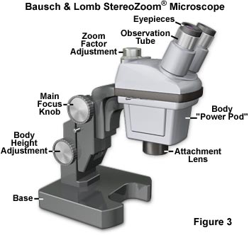
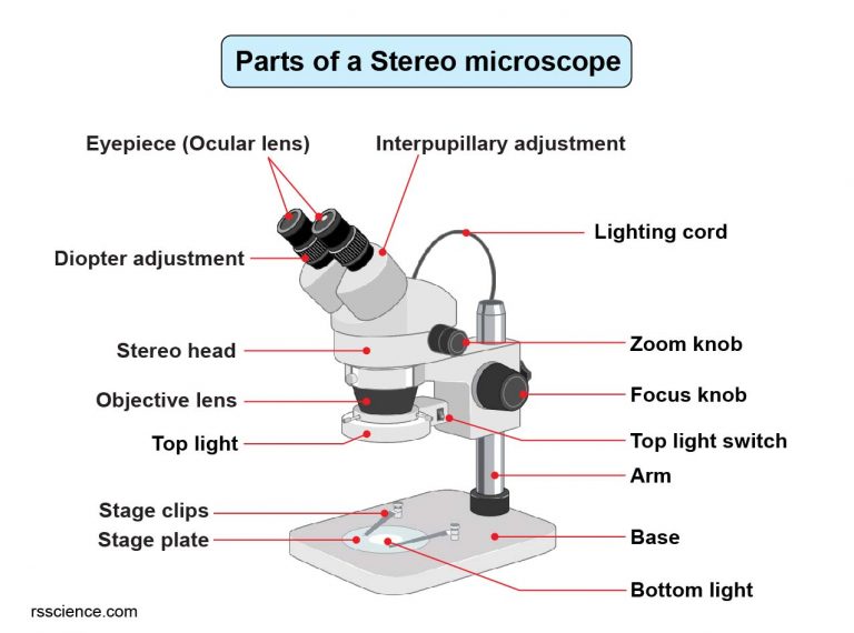
Oct 13, 2019 · Parts Of The Dissecting Microscope. Compound Microscope Parts Labeled. Microscope Diagram Labeled Unlabeled And Blank Parts Of A. Biology 2404. Simple Microscope Drawing At Paintingvalley Com Explore. Microscope Diaphragm. Compound And Stereo Microscopes Microscopes 4 Schools. Laboratory 5 The Microscope And Introduction To The Cell. Labeled Diagram of Dissecting microscope (Stereoscopic and Stereo microscope) A typical stereo microscope has 6 major parts which are:. LED Illuminators: Typically dissecting microscopes have an inbuilt LED light that acts a source of light in illuminating the specimen that needs to be observed. Eyepiece: Each dissecting microscope has two eyepieces that is used to focus on different pathways ... May 2, 2021 — The dissecting microscope has two magnifications known as Fixed magnification and Zoom magnification. Fixed magnification is used in eyepiece to ... A dissecting microscope has a single objective lens and two eyepiece lenses. Function: The compound microscope is used to observe minute and smaller things, like protozoa, bacteria, and cells, etc. It is used to study the surface of the specimen, in microsurgeries, for studying dissections, in watch and small circuit boards making, etc.
Samuel Broder, M.D., former Director, National Cancer Institute from 1989 to 1995.
Figure: Labeled Dissecting microscope (Stereo or stereoscopic microscope). Image created using biorender.com. LED illuminators-For some of the dissecting Microscopes, they have an inbuilt LED illuminator as a source of light.Eyepieces-They have two eyepieces each focusing different pathways of the light into and out of the specimen, each with its own magnification power.

Dissecting Microscope Labeled Diagram - Micropedia
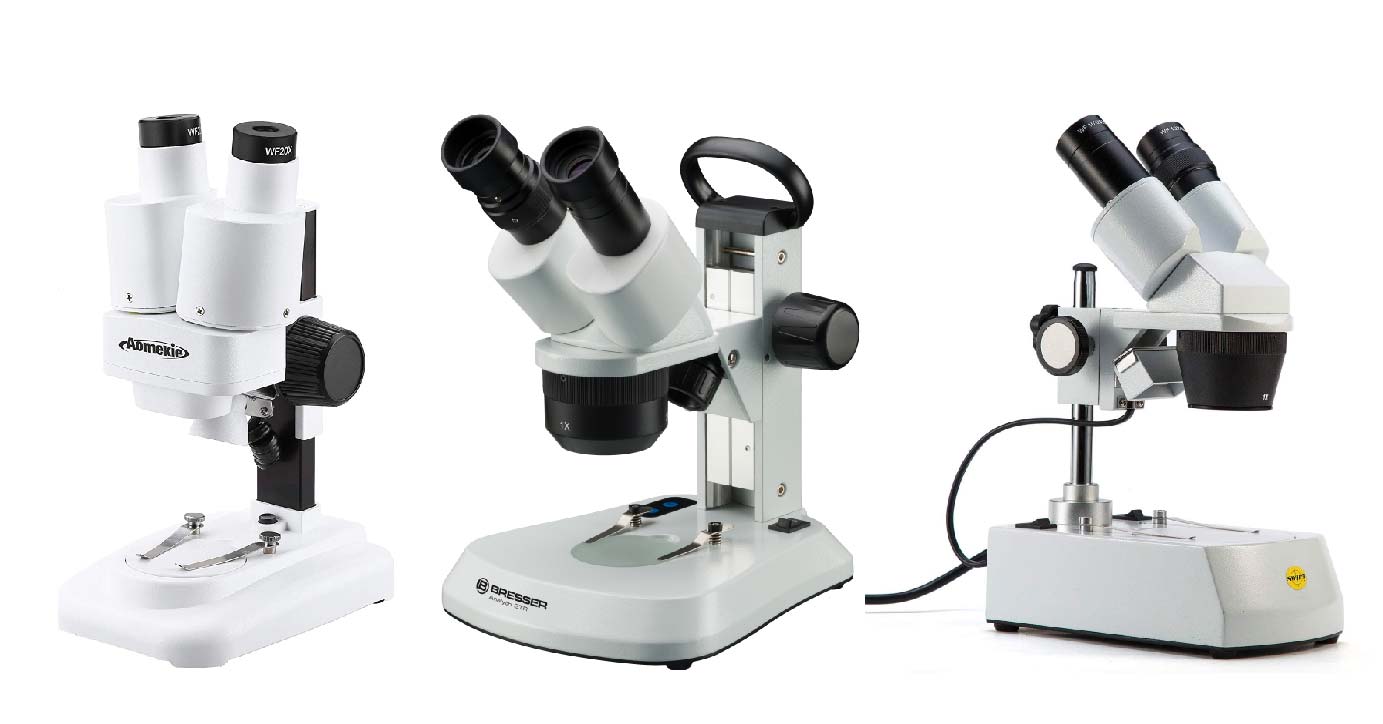
Stereo Microscope. The stereo microscope, also called a dissecting microscope, provides magnification of up to \(300\) times. These binocular microscopes are used to look at opaque objects or objects that are too large to be viewed with a compound microscope since they do not require a slide preparation.
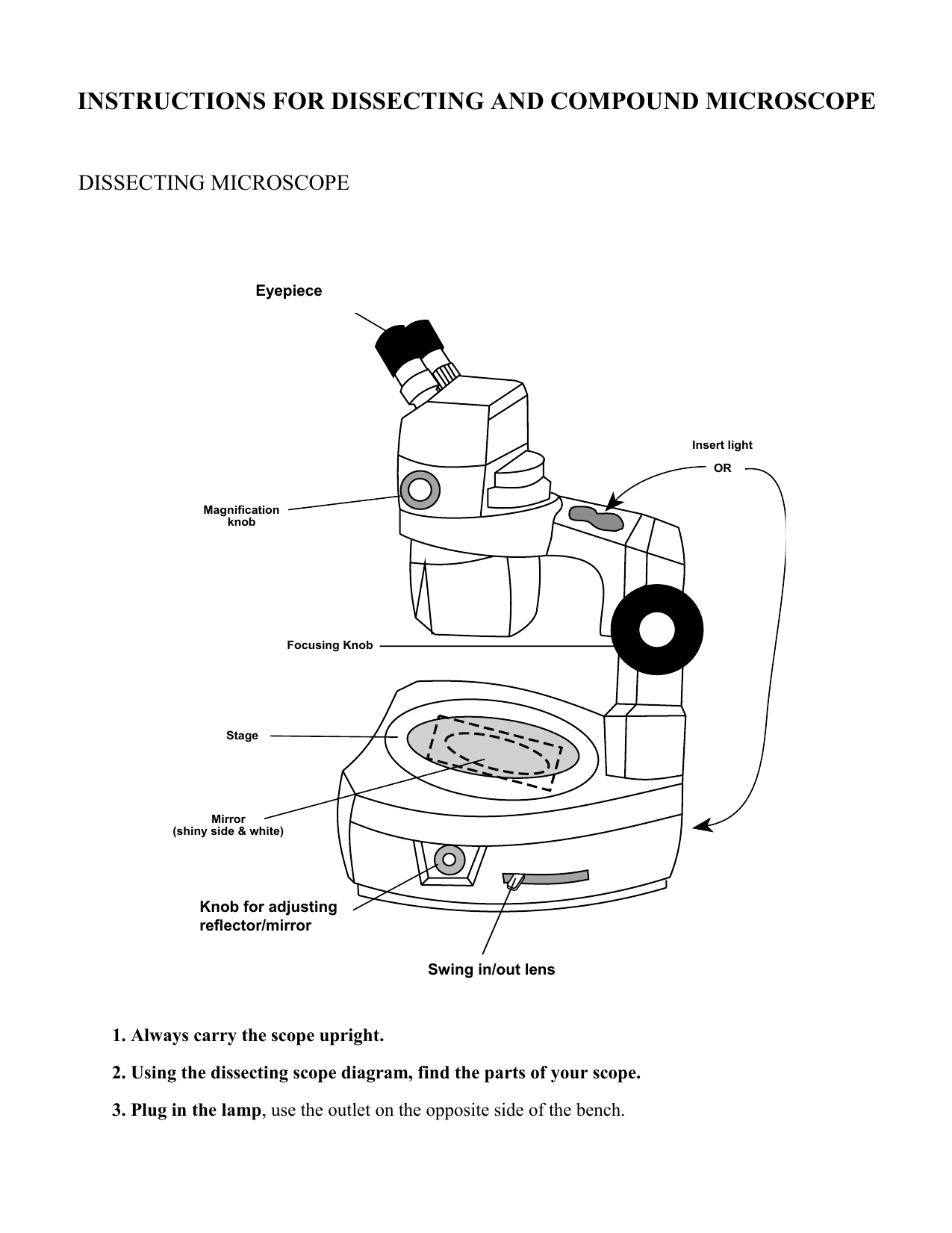
INSTRUCTIONS FOR DISSECTING AND COMPOUND MICROSCOPE ...
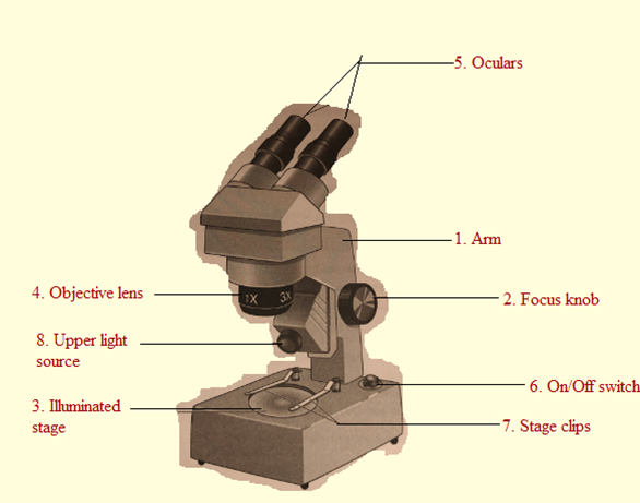
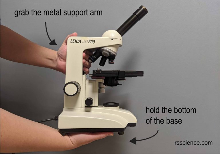
Compound Light. Used to examine material mounted on microscope slides (usually thinly sectioned & stained) Provides total magnification of 40x-1000x. No space for dissection. Rules. TRANSPORT. Arm & base. USE. Always start at 4x, Coarse focus, Fine focus.

Dissection Microscope at Rs 1500/piece | Dissecting ...
A Dissecting Microscope (also stereo microscope) on the other hand, has a longer working distance of up to 150mm and a lower magnification.A beam of light is projected from above the specimen. It is commonly used to view larger specimens and even perform dissections of small specimens such as insects.
Mycorrhizal Associations: Teaching Resources
Dissecting Stereo Microscope Parts and Functions Overview. Also known as a stereoscopic microscope, a dissecting microscope is a type of optical microscope commonly used for studying three-dimensional objects (3-D objects) as well as for dissecting biological specimen (e.g. insects and plant parts etc) at low magnification, between 2 and 100x depending on the microscope.
Answered: Microscopy 4.5 Use of the Dissecting… | bartleby
A Dissecting microscope is an optical microscope that allows the viewers to see a 3D image of the specimens. This microscope differs from the compound light microscope because they have a separate objective lenses and eyepieces.

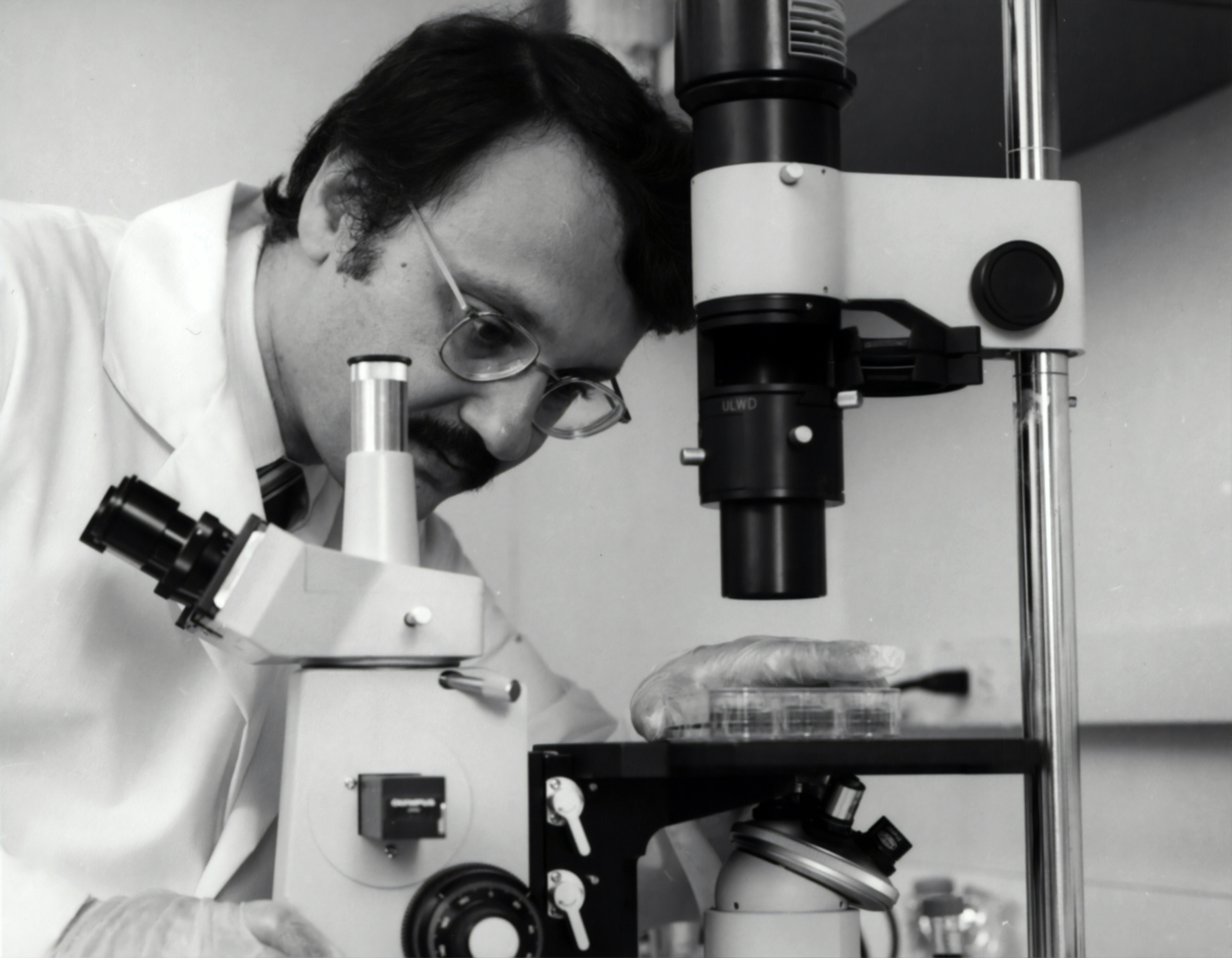
This Centers for Disease Control and Prevention (CDC) scientist was shown implementing molecular testing, in order to test for different types of polio. The 6-assay screening can determine which samples are polio, the specific serotype of polio, and whether they are vaccine, or wild strains.
A dissecting microscope, also known as a stereo microscope, is used to perform dissection of a specimen or sample. It simply gives the person doing the dissection a magnified, 3-dimensional view of the specimen or sample so more fine details can be visualized.

Dissecting Microscope Diagram | Clipart Panda - Free ...
Olympus Microscope Diagram. Here are a number of highest rated Olympus Microscope Diagram pictures upon internet. We identified it from well-behaved source. Its submitted by doling out in the best field. We allow this nice of Olympus Microscope Diagram graphic could possibly be the most trending topic next we allocation it in google pro or ...
This photograph depicted a Centers for Disease Control and Prevention (CDC) scientist, concentrating poliovirus from sewage, so that the virus could be grown in cultured cells, and then tested using molecular methods. She was performing a polio environmental surveillance technique. There is no routine polio environmental surveillance in the U.S., but surveillance is done in many countries.
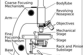
A simple (dissecting) microscope (Fig. 1.1) consists of the following parts. 1. Base - It is the basal part that is bifurcated and supports the weight of the microscope. It is generally horse-shoe shaped and is made of metal. 2. Stand - It is a short, hollow cylindrical rod fixed to the base. Another

Microscope Parts Diagram - Micropedia
A dissecting microscope, or more commonly known as a stereo microscope, is a microscope that gives a three-dimensional view of a specimen. This is because of the binocular head, or the two eyepieces that are slightly angled, which creates the perfect peripheral vision that results in a three-dimensional visual.
Parts of Stereo Microscope (Dissecting microscope ...
Simple/ Dissecting Microscope: As shown in the figure, dissecting microscope consists of a biconvex lens which is moved up and down by an adjustment screw to bring the object in sharp focus. The object is placed on the platform and light is focused with the help of a concave mirror fitted below.

Scanning Electron Microscope Blog: Fungi - Images for ...
Stereo microscopes are also called stereoscopic and dissecting microscopes.Compared to compound microscopes, dissecting microscopes have lower power optical systems and illuminators; a dissecting microscope's typical magnification ranges from about 10x-40x.Because stereo microscopes have less powerful magnification abilities, the types of objects or critters you can observe through them ...

Stereo microscopes are also | Clipart Panda - Free Clipart ...
In this article we will discuss about the parts of dissecting microscope with its working and utility. 1. Foot or Base: ADVERTISEMENTS: It is the basal, horse-shoe shaped or circular part of dissecting microscope. It is made of heavy material. It provides support to other parts of microscope.

Sketch Dissecting Microscope Diagram - Micropedia
5 Important Types Of Microscopes Used In Biology With Diagram. Solved 1 Familiarise Younelf With All The Parts Of The M. Stereo Microscope Basics. Microscope Parts Function Biological Science Picture Directory. Biology 2404. Http Www Lamission Edu Lifesciences Bio3labs Bio3 20lab03 Sp12 Microscopy 20 20cells Pdf.

Image from page 90 of "Text-book of structural and physiological botany" (1877)
A stereo microscope is defined as a type of microscope that provides a three-dimensional view of a specimen. It is also known as a dissecting microscope. In a stereo microscope, there are separate objective lenses and eyepiece such that there are two separate optical paths for each eye. Stereo Microscope Diagram. Principle of Stereo Microscope

Sketch Dissecting Microscope Diagram - Micropedia
Dissecting microscopes; Monocular, Binocular, and Trinocular microscopes; In addition to our selection of affordable microscopes for sale, we also offer a wealth of free resources. Find worksheets and experiments, a helpful diagram, a gift guide, a brief history of microscopes, and more.
Parts of Stereo Microscope (Dissecting microscope ...

Algae - Under the Microscope - YouTube

Image from page 20 of "Diagnosis and treatment of ear diseases" (1880)
Dissecting Microscope Labeled Diagram - Micropedia
Solved: 4.5 Use Of The Dissecting Microscope Label The Fol ...

Dissecting Microscope Diagram | Clipart Panda - Free ...

Binocular Microscope Labelled Diagram - Micropedia

Dissecting Microscope Field Of View - Micropedia

3AC---package-1

Micrographs made with a fluorescent dissecting microscope ...

Microscope - diagram Tom Butler | Science skills, Science ...
How to Use a Dissection Microscope

Closeup of skeleton hand model

Sketch Microscope Diagram Easy To Draw - Micropedia
This image depicted a Centers for Disease Control and Prevention (CDC) scientist interacting with her Caliper LifeSciences’ Zephyr Molecular Biology Workstation, working with samples to be tested using a real-time PCR machine, known as a themocycler (see PHIL 22904), in order to identify the various types of poliovirus contained therein. The data from this analysis is stored in a computer, while the software further analyzes the data before being reviewed by a scientist.
Robert Charles Gallo, former Biomedical Researcher. He is best known for his work with the Human Immunodeficiency Virus (HIV), the infectious agent responsible for the Acquired Immune Deficiency Syndrome (AIDS). He was the former Chief of Laboratory of Tumor Cell Biology at the National Institutes of Health. 1980
Simple Parts Of A Microscope Diagram - Micropedia

Mr. Klein's Classes - science_handbook_4.3 | Science ...
Dissecting Microscope Labeled Diagram - Micropedia
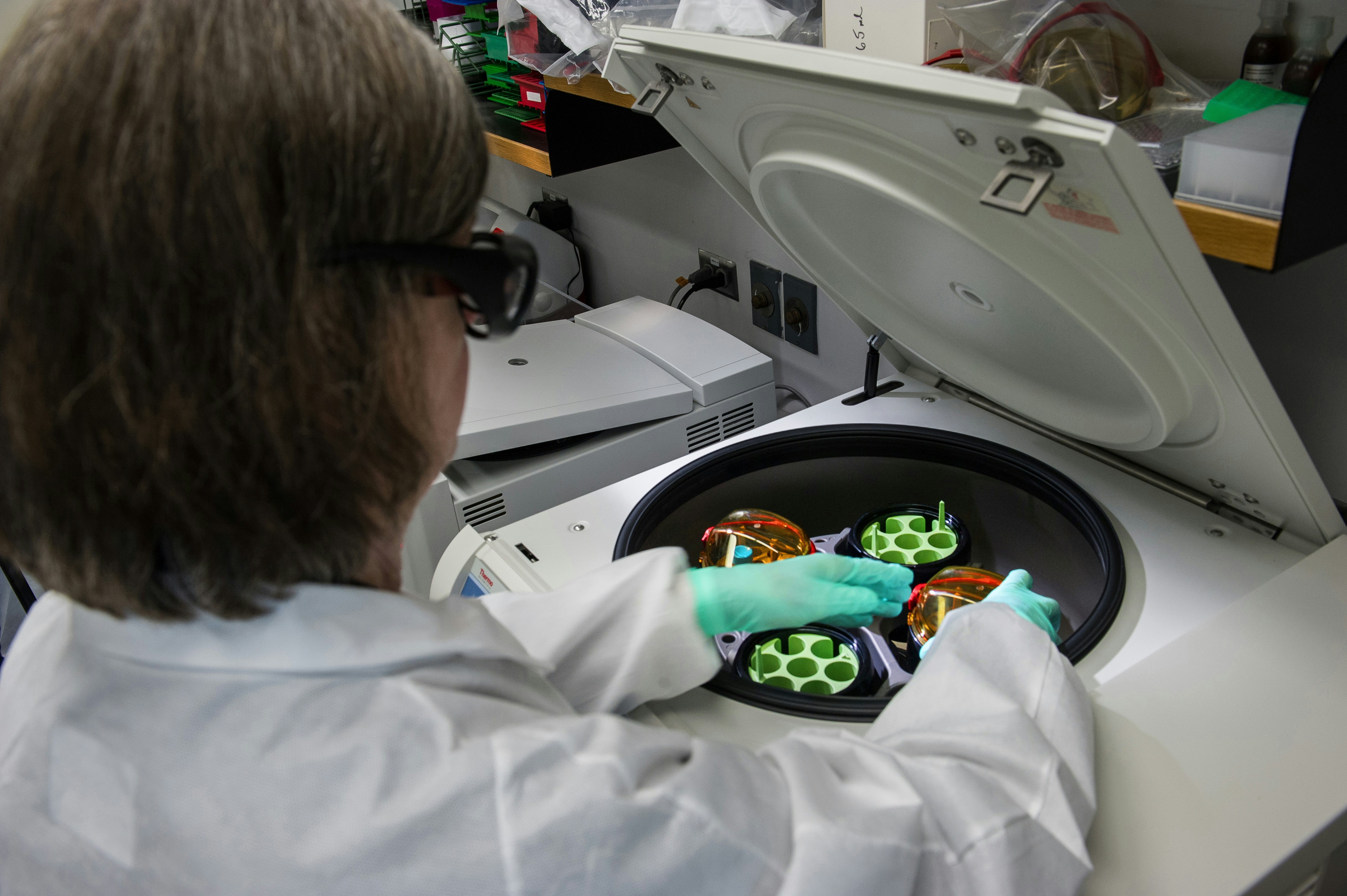
This Centers for Disease Control and Prevention (CDC) scientist was shown working with stool samples, which had been mixed with chemicals, and was placing the chemical mixtures into a centrifuge, which when spun up to a high rate of speed, would separate the solid material from the liquid supernatant.

Image from page 265 of "Technology of textile design. Being a practical treatise on the construction and application of weaves for all textile fabrics, with minute reference to the latest inventions for weaving. Containing also an appendix showing the ana
Parts of Stereo Microscope (Dissecting microscope ...
Microscopes Drawing at GetDrawings | Free download

Choosing a Microscope | Make: DIY Projects and Ideas for ...

CELLS: ORIGINS


Comments
Post a Comment