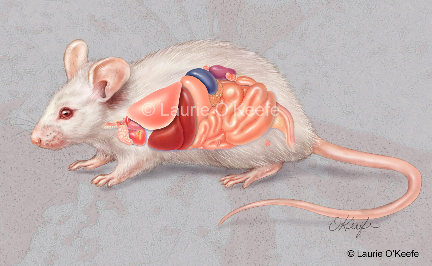
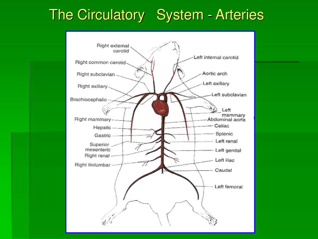
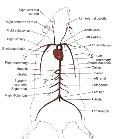
42 mouse dissection diagram
PROSECTION OF THE MOUSE - University of South Florida Diagram Key • 1. Superficial cervical nodes • 2. Deep cervical nodes • 3. Mediastinal nodes • 4. Axillary node • 5. Brachial node • A. Thymus • B. Spleen • 6. Pancreatic node • 7. Renal nodes • 8. Mesenteric node • 9. Inguinal node • 10. Lumbar nodes • 11. Sacral Node • 12. Sciatic node DOCX RAT DISSECTION - Eagle Mountain-Saginaw Independent School ... Therefore, please follow the instructions outlined in this lab for proper dissection technique and never cut more than is absolutely necessary to expose an organ. Raise structures that you wish to cut with forceps so that you can see what lies underneath. Approach the dissection in a step-like manner.
PDF Field Mouse Dissection - Continued - biology with mrs. h & Day 2: Internal Anatomy During$this$portion$of$the$dissection,$draw$and$label$each$bolded$itemonthemousediagrambelow. $ 1.

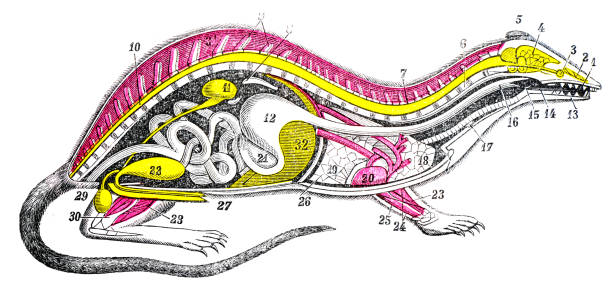
Mouse dissection diagram
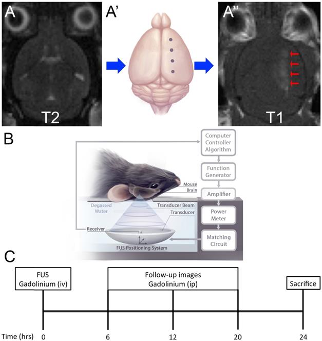
Dissection of the ventral midbrain region from mice ... Dissection of the ventral midbrain region from mice. Bregma coordinates and anatomic boundaries were established for dissection of ventral midbrain based on the mouse brain atlas (42). Briefly, the... PDF Mouse dissection - people.springfield.k12.or.us The mouse is left-right as diagramed in Figure 1. Figure 1 2. Limbs (Left forepaw) Observe the paws of the mouse. In Figure 2 you will see the layout of these. See if you can identify the parts of the paw. 3. Determine the gender of your animal: Using the diagrams below, look at your mouse and circle which gender your mouse is. Mouse Anatomy Mouse Anatomy Anatomy Quicktime movies of Histologic Anatomy HEART/LUNG KIDNEY MAMMARY GLAND LYMPH NODE PROSTATE SPLEEN LIVER SALIVARY GLANDS 3-D WIRE MODEL BASED ON MRI SECTIONS Quicktime Mouse Radiographic Atlas of Skeletal Anatomy The following link will take you to a series of radiographic images with color overlays and labels.
Mouse dissection diagram. Mouse Brain Gross Anatomy Atlas - MBL Mouse Brain Gross Anatomy Atlas : Home Brain Atlases iScope Brain Library MBL Procedures Databases Movies: Links : Mouse Brain Gross Anatomy Atlas [Part I Fixed Brain][Part II Fresh Brain] 1-4 5-8 9-12 13-16 17-20 21-24 25-28 29-32 33-36 : Step5 : Case ID: 063099.18 : Body Weight: 19 g ... PDF RAT DISSECTION GUIDE - philipdarrenjones.com The diagram below illustrates the muscles of the ventral surface of the rat. Be able to identify those listed. Use the photographs on the following pages and your lab atlas to assist you. Head and Throat muscles Digastric - this V shaped muscle follows the lower jaw line. It functions to open the mouth. Mouse Lymph Node Dissection - development of a unique ... Here are a number of highest rated Mouse Lymph Node Dissection pictures upon internet. We identified it from obedient source. Its submitted by meting out in the best field. We acknowledge this kind of Mouse Lymph Node Dissection graphic could possibly be the most trending topic in the same way as we part it in google gain or facebook. Mouse Anatomy Diagram | Worksheet | Education.com Mouse Anatomy Diagram Can your preschooler name the parts of a mouse? Give her a dose of animal science with this fun cut-and-paste printable. Help her complete the mouse chart by showing her how to cut out the names of the parts and paste them in the right spaces.
PDF Field Mouse Dissection - biology with mrs. h The Anatomy of the Laboratory Mouse 10. Skeleton of LAC Grey mouse. 11. Dorsal aspect of skull. 12. Left lateral aspect of skull. 13. Basal aspect of skull. 14. Lateral aspect of right mandible. 15. Medial aspect of right mandible. 16. Atlas (1st cervical vertebra). Anterior aspect (above). Posterior aspect (below). 17. Axis (2nd cervical vertebra). Anterior aspect (above). C:\BIOL198\mouse dissection.wpd The mouse is left-right as diagramed in Figure 1. 2. First, pin the animal down with his/her belly facing up. With ethanol, wet the animal down. By washing the carcass with ethanol, you are protecting the tissues from artifacts caused by hair dragging through them. 3. With your forceps, grab hold of the skin anteriorly to the urethral opening. Procedure of dissection muscles from a mouse. A. Tools ... Context 1 ... Cut the tendon of gastrocnemius (GA) muscle and use it to peel off the GA muscle till the exposure of a SOL tendon ( Figure ... View in full-text Context 2 ... Cut the SOL tendon and...
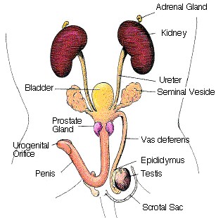
rat dissection - Evolving Sciences A rat dissection is a perfect way to show all the organs in the body. You can analyse the circulatory system, respiratory system and digestive system through this dissection. As you are dealing with a organisms make sure you clean your benches after use, use gloves and a lab coat. Take the appropriate safety steps when dealing with the ... The Anatomy of the Laboratory Mouse Margaret J. Cook urogenital system. 65. Male reproductive organs. 66. Right testis. 67. Bladder and prostate. 68. Female urogenital system.1. Latex injection apparatus: 2. Latex injection ... Virtual Rat Dissection Step by Step - The Biology Corner Rat External Anatomy. Step 1: In the biology lab, you will be working with specimens that have been preserved in chemicals and you will be working with sharp instruments. Before you start, obtain safety goggles, and nitrile gloves. Nitrile gloves come in different sizes, most women will wear a medium and most men will wear a large. PDF Laboratory Detectives — Owl Pellet Dissection 101 Chart of Mouse, Vole or Rat Skeletons Glue PROCEDURE 1. Have students wear plastic, disposable gloves. Remember you are dissecting something that was once inside an owl! 2. Measure and note the shape of the pellet by tracing or drawing its shape. Ask students if they think this pellet came from a large or small owl. 3.
PDF Mouse dissection Kelley - mbl.edu This series of diagrams is intended to serve as a guide for the cochlear dissection. Pinning: 1. Align temporal bone so that the cochlea is pointing towards the back of the dish and the vestibular portion is towards the front. 2.
Anatomy of the laboratory mouse: in vivo imaging atlas on ... Labeled cross-sectional anatomy of the mouse on micro-CT. These images of a normal female Swiss mouse have been acquired with a laboratory-based microCT system (nanoScan PET/CT Mediso (Budapest, Hungary) with an operation voltage of 50kVp and a 0,14mm pitch, with an intravenous injection of 2ml of Visipaque (320mg d'I/ml), at CERIMED (Centre Européen de Recherche en Imagerie Médicale ...
Lymph node mapping in the mouse Accurate identification of lymph nodes in the mouse is critical for studies of tumor metastasis, and of regional immune responses following immunization. However, these small lymphatic organs are often difficult to identify in mice using standard dissection techniques, so that larger rats have been …
Why Do People Craze For These 10 Mouse Anatomy Diagram ... Finding the mouse anatomy diagram review is somewhat daunting when you have plenty of choices to pick. Many of us are getting frustrated especially when we are first-time consumers of mouse anatomy diagram. Here we will discuss different types of choices with unique features of them and give you a broad outline to choose between […]
Parts of a Mouse Diagram - Quizlet When the screen pointer is over the icon of the object, the mouse button is clicked to grab it then you drag it to its new location. Drag and Drop a gesture in which the user selects a virtual object by Clicking it and dragging it to a different location or onto another virtual object.
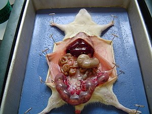
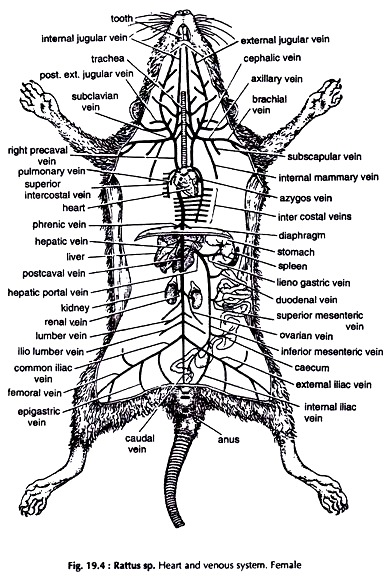
Dissection of Rat (With Diagram) | Zoology Dissection: Put the specimen on its back on a dissecting tray. Fix it with pins passing through the limbs. Lift the skin of the abdomen with a pair of forceps and make a small cut at about the middle of the abdomen. Starting from the cut give an incision extending up to the snout anteriorly and the genital opening posteriorly.
Cellular anatomy of the mouse primary motor cortex | Nature Cellular anatomy of the mouse primary motor cortex ... type-based wiring diagram of the mouse MOp-ul that will facilitate future analyses of motor control infrastructure across molecular, cellular ...
PDF YOUR GUIDE TO DISSECTION - Philip Harris Ltd. YOUR GUIDE TO DISSECTION Since 1817 Dissecting an Eye Before dissection, allow the students to a have a good look at the Eye and see if they can identify any parts of the Eye. Before commencing any kind of dissection on animal material, always read and implement any Health & Safety measures.
Mouse Anatomy - AALAS Illustrations on the mouse anatomy and rat anatomy are provided for the following: Body Regions Juvenile Features and Sex Differentiation External Features Mammary Glands Structures of the Head and Neck Heart, Vascular Tree, and Respiratory Tract Abdominal Structures Male Urogenital Apparatus Female Urogenital Apparatus
Dissection of E7.5 mouse embryos for whole embryo culture ... Transfer all conceptuses to a fresh dish of dissecting ... View in full-text Context 5 ... starting at the anti-mesometrial (fluffy) end (Fig. 1B), to reveal the trophoblast layer which envelops...
Mouse dissection - YouTube How does a mammal breath? How its reproductive system is done? «Mouse dissection» is a scientific movie and a pedagogic document for Biology students. The HD...
Rat Dissection Guide - VWR 1. Illustrated by Veronica Zoeckler. © 2015 Ward's Science All Rights Reserved. Page 4. Rib cage. Lung. Heart. Diaphragm. Liver. Stomach. Spleen. Small.16 pages
Mouse Anatomy Mouse Anatomy Anatomy Quicktime movies of Histologic Anatomy HEART/LUNG KIDNEY MAMMARY GLAND LYMPH NODE PROSTATE SPLEEN LIVER SALIVARY GLANDS 3-D WIRE MODEL BASED ON MRI SECTIONS Quicktime Mouse Radiographic Atlas of Skeletal Anatomy The following link will take you to a series of radiographic images with color overlays and labels.
PDF Mouse dissection - people.springfield.k12.or.us The mouse is left-right as diagramed in Figure 1. Figure 1 2. Limbs (Left forepaw) Observe the paws of the mouse. In Figure 2 you will see the layout of these. See if you can identify the parts of the paw. 3. Determine the gender of your animal: Using the diagrams below, look at your mouse and circle which gender your mouse is.
Dissection of the ventral midbrain region from mice ... Dissection of the ventral midbrain region from mice. Bregma coordinates and anatomic boundaries were established for dissection of ventral midbrain based on the mouse brain atlas (42). Briefly, the...








![Mouse Dissection || Of Mice and Men [EDU]](https://i.ytimg.com/vi/RRs59csAQws/maxresdefault.jpg)

Comments
Post a Comment